
Case: 43- year old male with 1 cm painless lesion in the oral cavity
Histopathology

Case: 43- year old male with 1 cm painless lesion in the oral cavity

pathologymcqs2 min read
Case discussion: oral cavity lesion in a 43-year-old maleCase reports of pseudoepitheliomatous hyperplasiaCase study: 1 cm lesion in the oral cavityCase study: pseudoepitheliomatous hyperplasia in oral cavityClinical approach to painless oral lesionsClinical approach to pseudoepitheliomatous hyperplasiaCommon causes of oral lesions in adultsDiagnosis and management of pseudoepitheliomatous hyperplasiaDiagnosis of painless oral lesions in middle-aged malesDifferential diagnosis of pseudoepitheliomatous hyperplasiaDifferentiating benign hyperplasia from malignant lesionsDifferentiating pseudoepitheliomatous hyperplasia from SCCEvaluating oral cavity lesions in middle-aged menHistological features of pseudoepitheliomatous hyperplasiaHistopathological differentials for pseudoepitheliomatous hyperplasiaHistopathological examination of oral lesionsHistopathology of pseudoepitheliomatous hyperplasiaIdentifying benign and malignant oral lesionsIdentifying painless lesions in the oral cavityIdentifying pseudoepitheliomatous hyperplasia in clinical practiceManagement of painless lesions in the mouthMiddle-aged man with painless oral lesion case reportOral lesion biopsy results: case studiesOral pathology case study: painless lesionsOral pathology: painless lesions in adultsPainless lesions in the mouth: what to look forPainless oral cavity lesions differential diagnosisPainless oral lesions: clinical presentation and diagnosisPathology of oral cavity lesions in adultsPseudoepitheliomatous hyperplasia clinical featuresPseudoepitheliomatous hyperplasia diagnosisPseudoepitheliomatous hyperplasia treatment optionsPseudoepitheliomatous hyperplasia vs. squamous cell carcinoma in biopsySquamous cell carcinoma vs. pseudoepitheliomatous hyperplasiaSymptoms and diagnosis of oral cavity lesionsUnderstanding painless oral cavity growthsUnderstanding pseudoepitheliomatous hyperplasia in oral pathology

Watch the video completely

https://youtube.com/shorts/86YfcIEJJFo?feature=shared

How to differentiate between squamous cell carcinoma and pseudoepitheliomatous hyperplasia (PEH)?
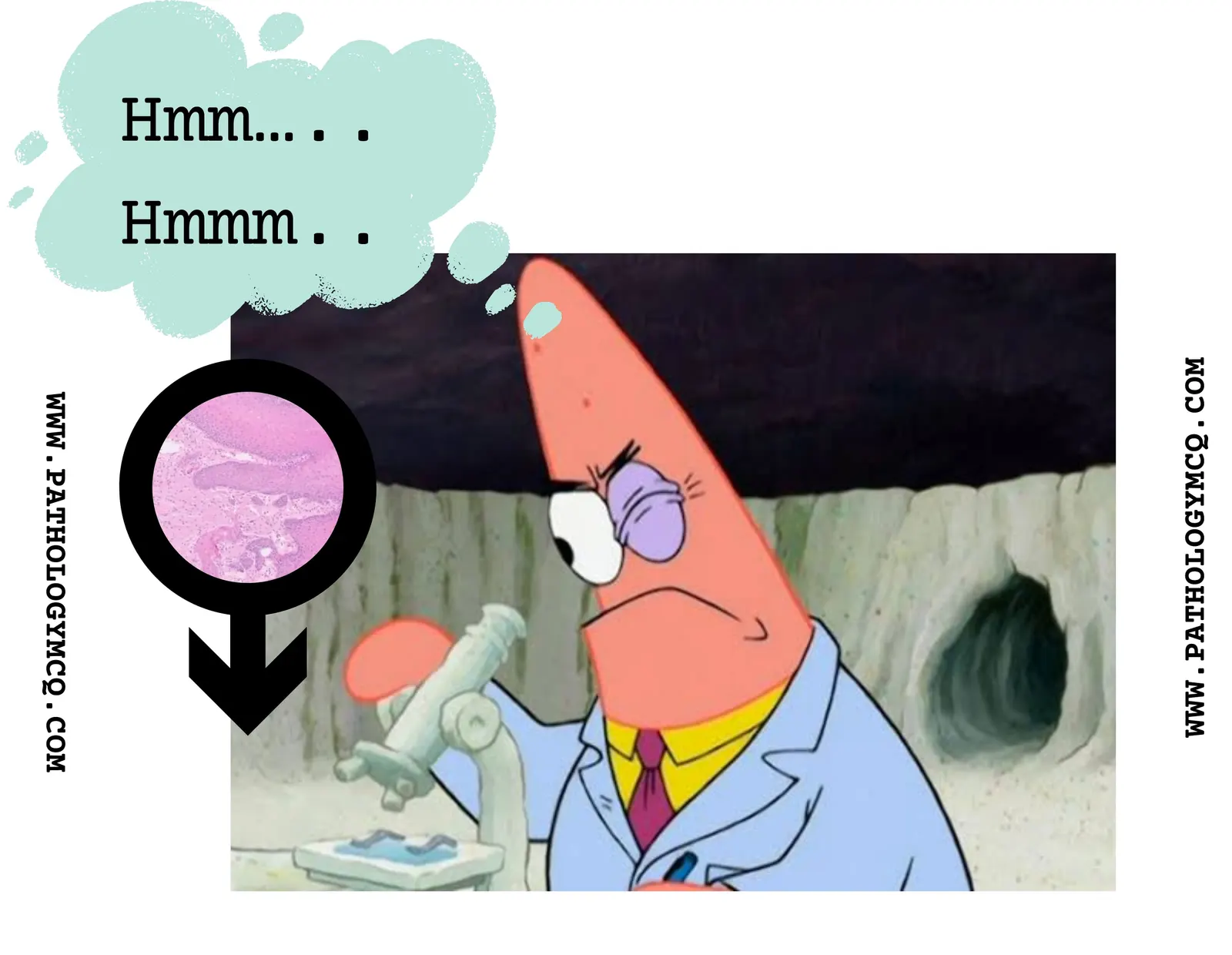
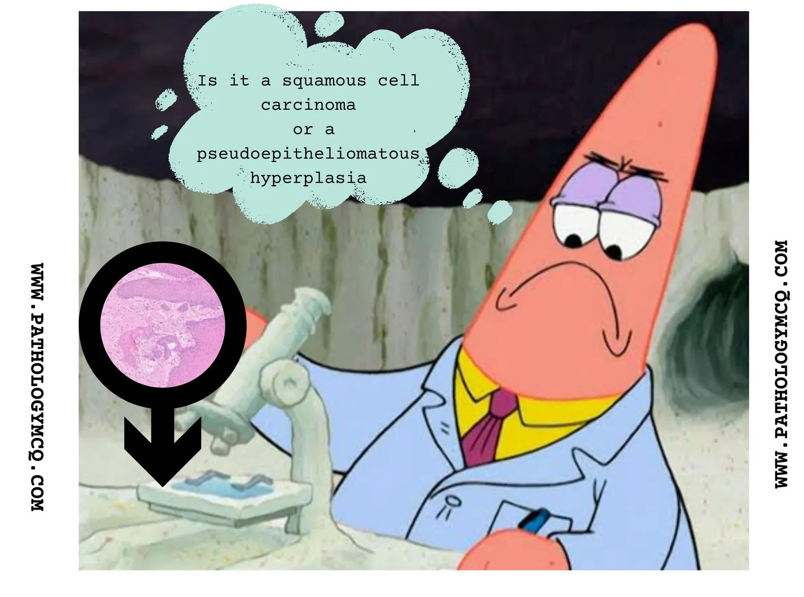
Questions to ask:

  • Is the orientation proper- if not, consider reorienting.
  • Is proper clinical history available- get clinical history especially in difficult cases

Suspect pseudo-epitheliomatous hyperplasia when

  1. You see elongation of epithelium, interconnected rete-ridges (these may simulate pseudo invasion) In the absence of significant atypia
  2. When prominent lymphocytic or neutrophilic inflammatory infiltrates are identified. (suspect infectious etiologic in these cases)
Image: Keratin pearls identified in the dermis without significant atypia: Pseudoepitheliomatous hyperplasia in granular cell tumor.

Role of IHC: Studied to distinguish between invasion and pseudo invasion. p53 shows diffuse nuclear positivity in tumor cells (true invasion) whereas negative in PEH. E-cadherin is lost in tumor cells whereas intact in PEH.

Conditions associated with PEH.: Granular cell tumor, Candidiasis (median rhomboid glossitis, chronic hyperplastic candidiasis), Necrotizing sialometaplasia , Deep chronic infections, such as blastomycosis , Granulomatosis with polyangiitis , Oral submucous fibrosis, Melanocytic nevi, Melanoma.

Do add your own tips in the comments below.

References:

  • Cooper, P. H. (2005). Distinguishing Pseudoepitheliomatous Hyperplasia from Squamous Cell Carcinoma in Cutaneous Lesions. Archives of Pathology & Laboratory Medicine, 129(8), 1032-1036. https://doi.org/10.5858/2005-129-1032-DPHFSC
  • Kodet, O., Lacina, L., Krejčí, E., Dvořánková, B., Smetana, K., & Matoušková, E. (2015). Pseudoepitheliomatous hyperplasia: Review. International Journal of Clinical and Experimental Pathology, 8(7), 8580-8585. https://www.ncbi.nlm.nih.gov/pmc/articles/PMC4589711/
  • Santoro, A., Pannone, G., Contaldo, M., Pagliuca, M. Q., Serpico, R., & Maiorano, E. (2015). Pseudoepitheliomatous hyperplasia: A misleading mimic of squamous cell carcinoma. International Journal of Surgical Pathology, 23(2), 110-115. https://doi.org/10.1177/1066896914566834

For more such diagnostic tips- JOIN US in our approach based course

/blog/product/approach-based-course-histopathology-and-cytology/

Related articles