
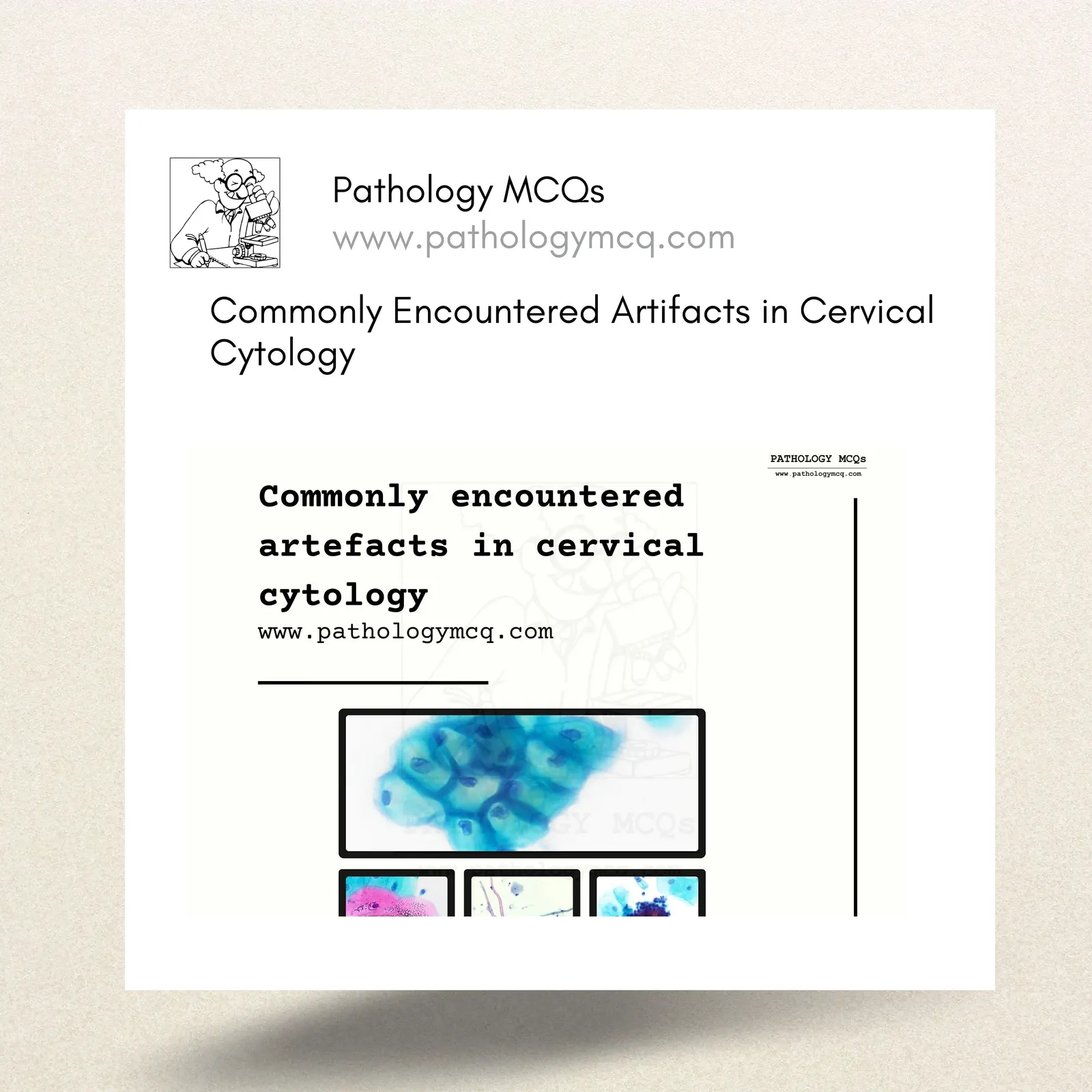
Cervical cytology plays a crucial role in diagnosing various gynecologic conditions, especially in screening for cervical cancer. However, artifacts in cytology slides can sometimes complicate interpretations, leading to potential misdiagnosis. Here, we explore some of the commonly encountered artifacts in cervical cytology, which every pathologist should be aware of.
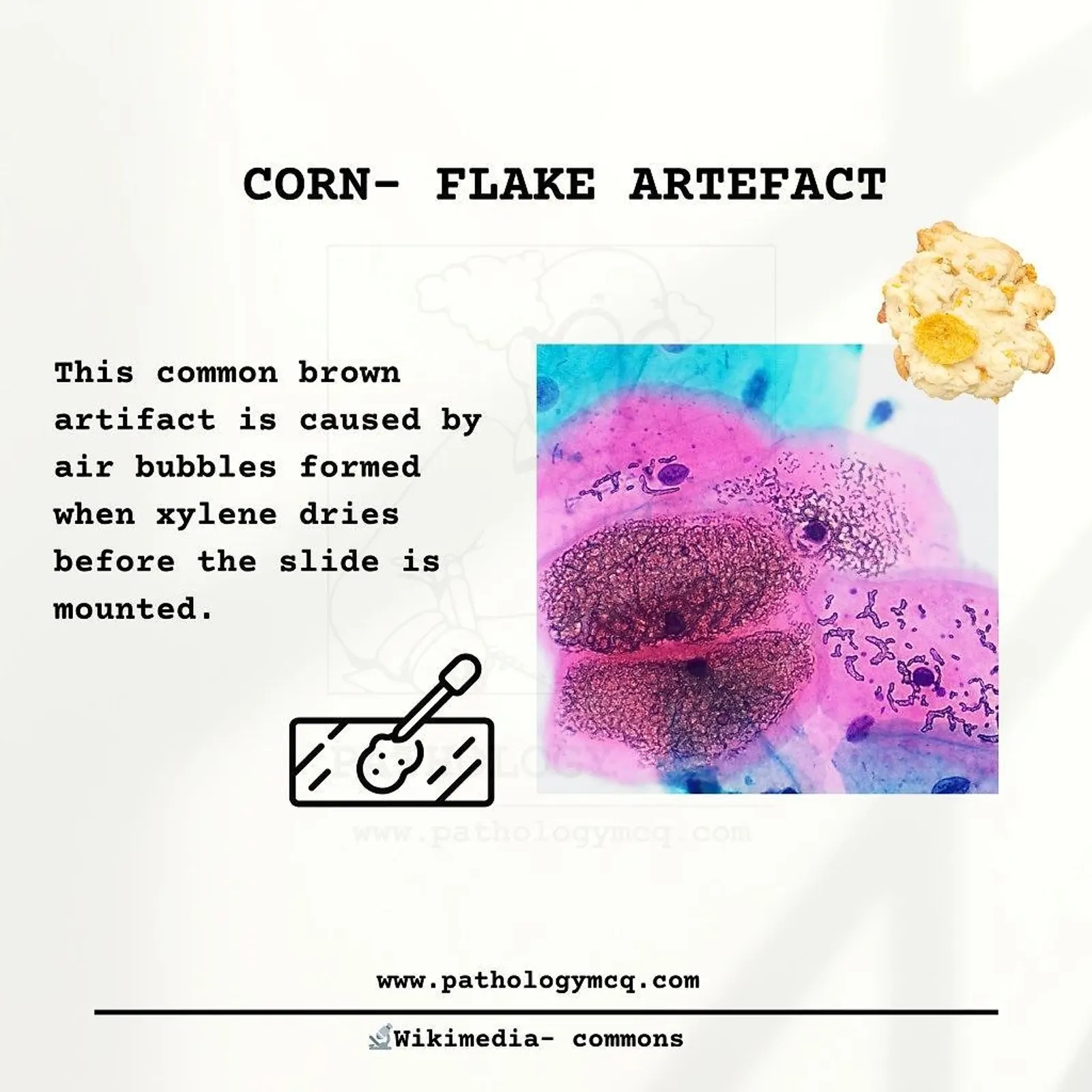
1. Cornflake Artifact
This common brown artifact appears due to air bubbles that form when xylene dries before the slide is mounted. Named for its resemblance to cornflakes, this artifact can be mistaken for pathological findings if not identified correctly. It highlights the importance of proper slide preparation and mounting techniques to avoid diagnostic errors.
Cause: Air bubbles due to drying of xylene before mounting
Appearance: Brown, flaky structures on the slide
Preventive Tip: Ensure slides are adequately mounted to avoid air bubble formation.
2. Cocklebur Artifact
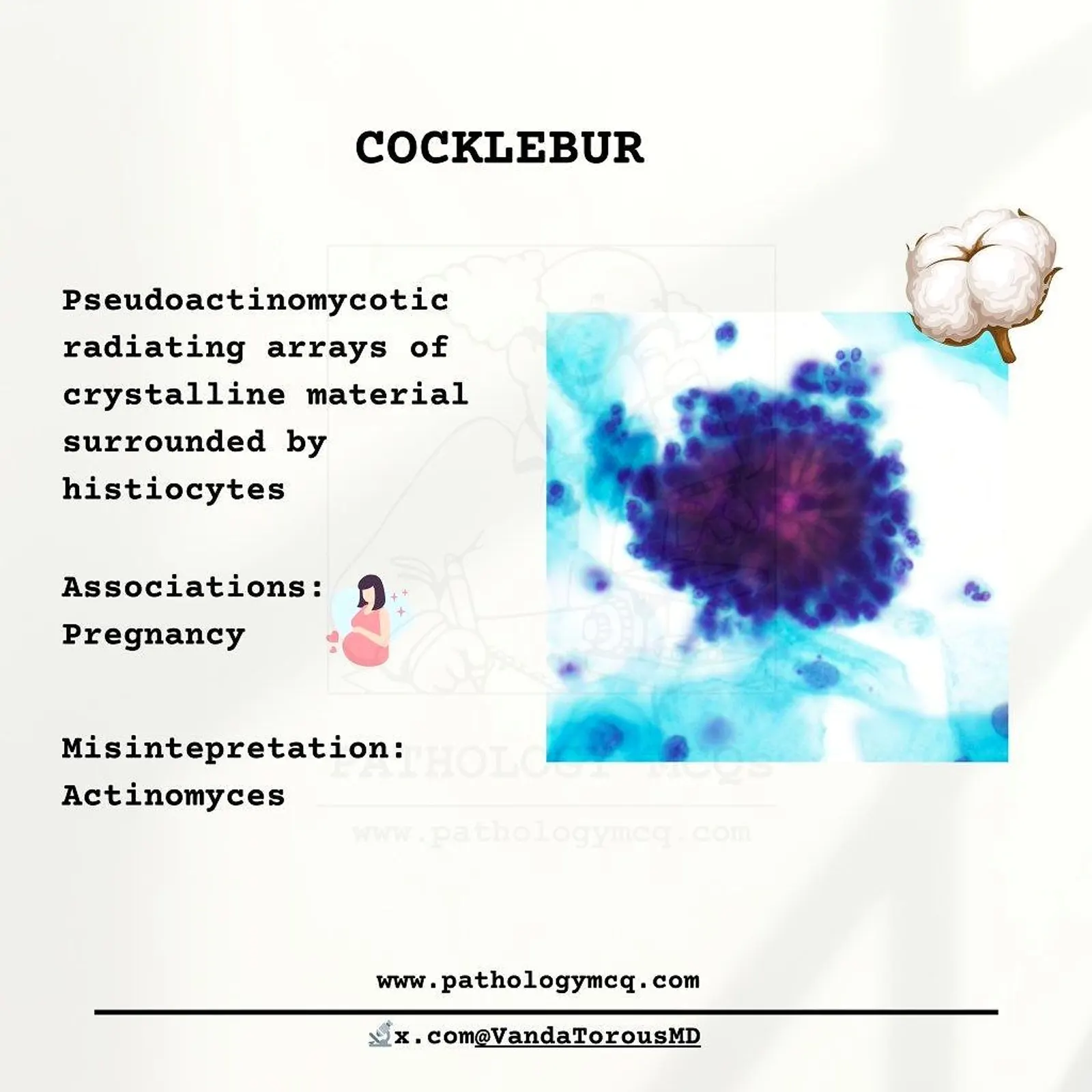
Cocklebur artifacts, also known as pseudoactinomycotic radiating arrays, are crystalline material surrounded by histiocytes. They are sometimes associated with pregnancy and can be mistaken for Actinomyces infection due to their similar radiating appearance.
Associations: Often seen in pregnancy
Misinterpretation: Can be confused with Actinomyces
Identification Tip: Note the radiating crystalline structure and absence of true fungal elements.
3. Glycogenation
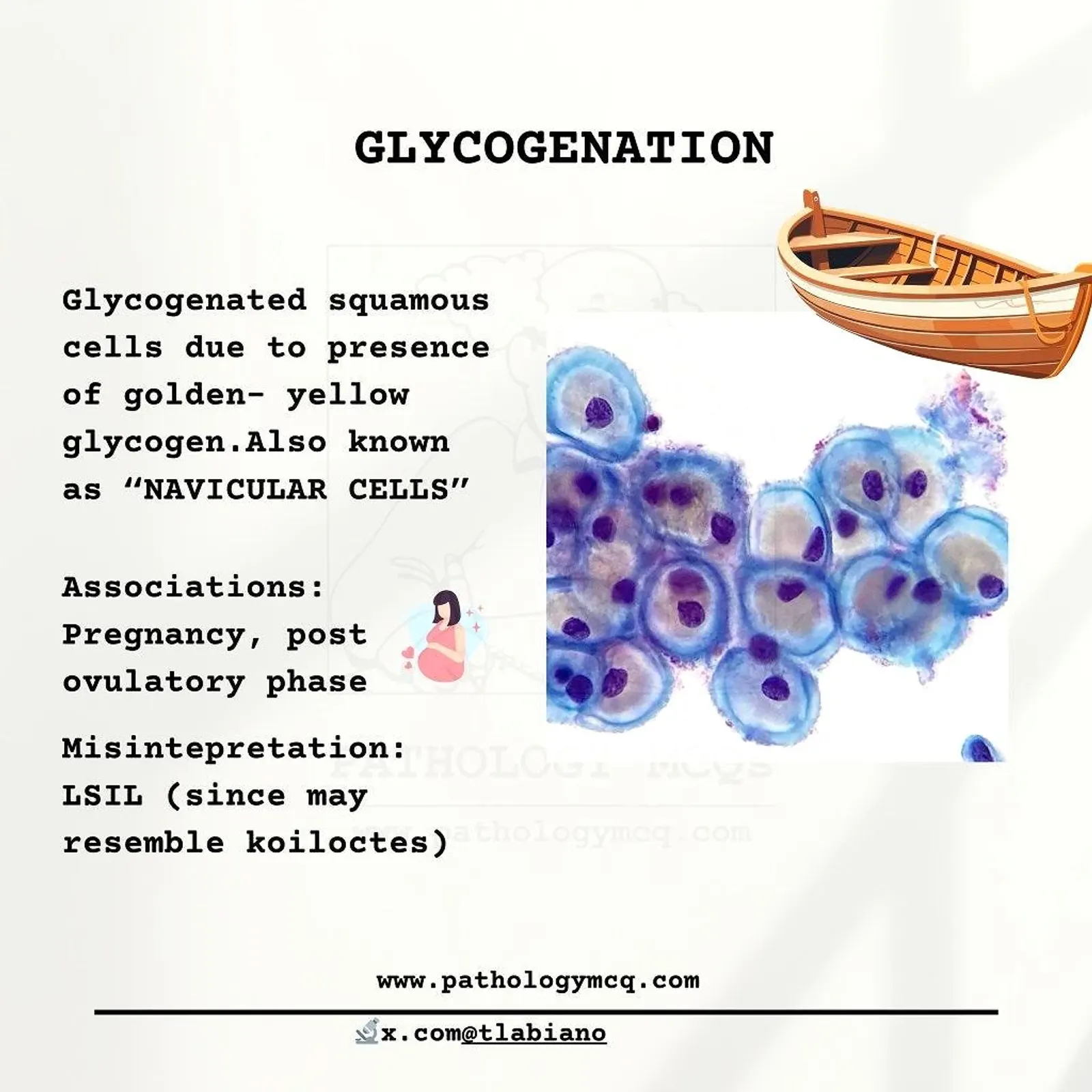
Glycogenated squamous cells, also called “navicular cells,” display a golden-yellow hue due to glycogen accumulation. This phenomenon is typically associated with pregnancy and the post-ovulatory phase. In some cases, glycogenated cells may resemble low-grade squamous intraepithelial lesion (LSIL), leading to possible misinterpretation.
Associations: Pregnancy, post-ovulatory phase
Misinterpretation: May resemble LSIL due to similar cellular changes
Identification Tip: Look for the golden-yellow glycogen deposits in the cells.
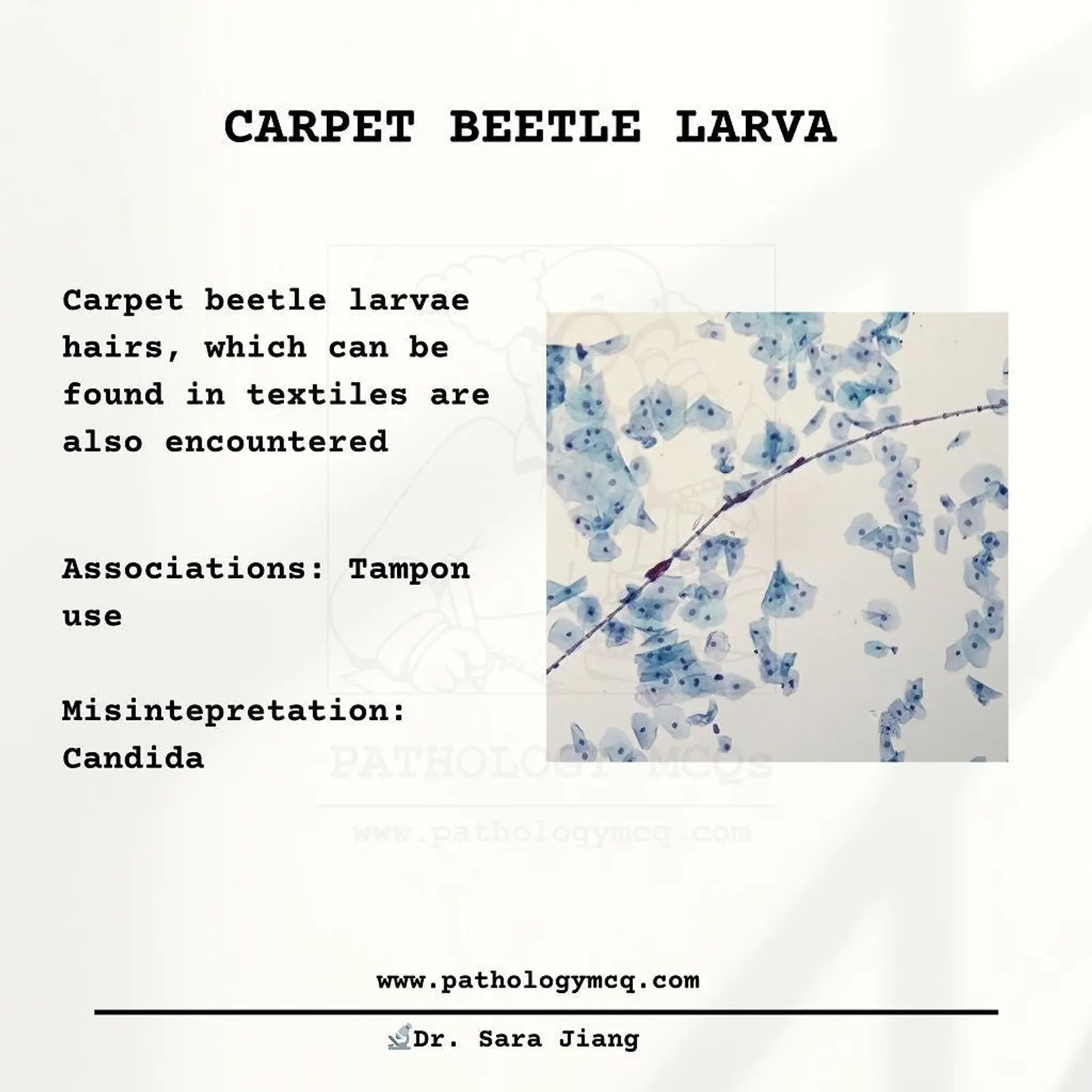
4. Carpet Beetle Larva
Carpet beetle larvae hairs, commonly found in textiles, can sometimes appear on cytology slides, especially in cases involving tampon use. This artifact can be mistaken for fungal infections, such as Candida.
Associations: Tampon use
Misinterpretation: Often misinterpreted as Candida
Identification Tip: Observe the characteristic hair-like structures to differentiate it from fungal elements.
5. Trichromes
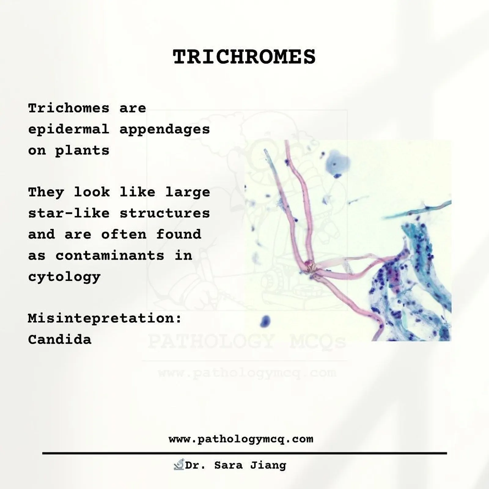
Trichromes are plant-derived contaminants that appear as large, star-like structures on cytology slides. These structures, often found in the context of plant exposure, can be mistaken for Candida infections due to their unique morphology.
Cause: Plant epidermal appendages
Appearance: Large, star-like structures
Misinterpretation: Can be confused with Candida
Identification Tip: Recognize the characteristic structure to avoid confusion with fungal organisms.
For more like these: GET OUR CYTOPATHOLOGY NOTES

References
Cibas, E. S., & Ducatman, B. S. (2014). Cytology: Diagnostic Principles and Clinical Correlates (4th ed.). Philadelphia, PA: Elsevier Saunders.
Mitchell, M. L., & Medley, R. A. (2013). “Recognition of Artifacts and Contaminants in Cervical Cytology: A Practical Approach.” Journal of Cytology, 30(1), 27-32