
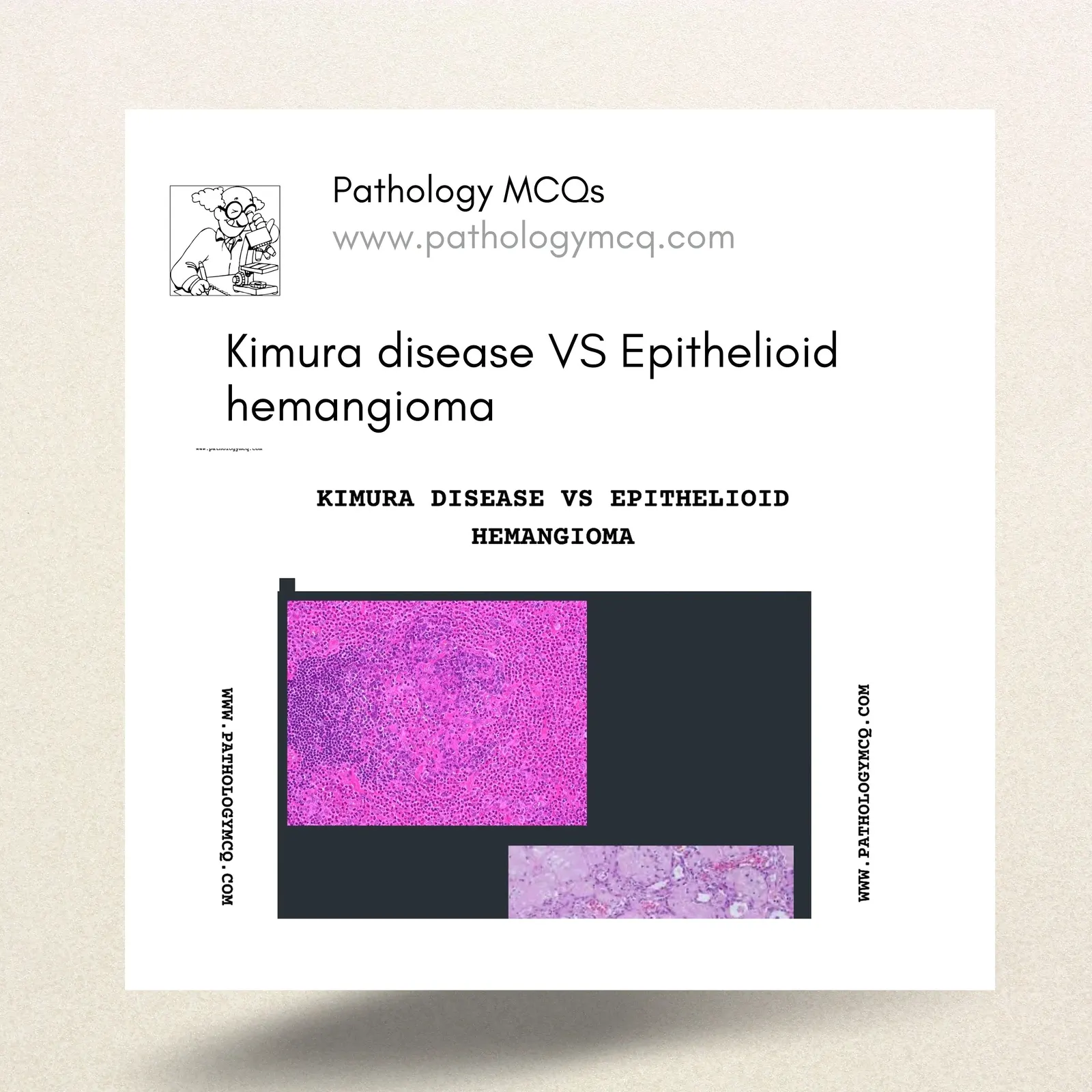
Kimura disease and Epithelioid hemangioma (Previously , Angiolymphoid hyperplasia with eosinophilia (ALHE) ) are often confused and need to be differentiated. Here are a few differences.
1.AGE
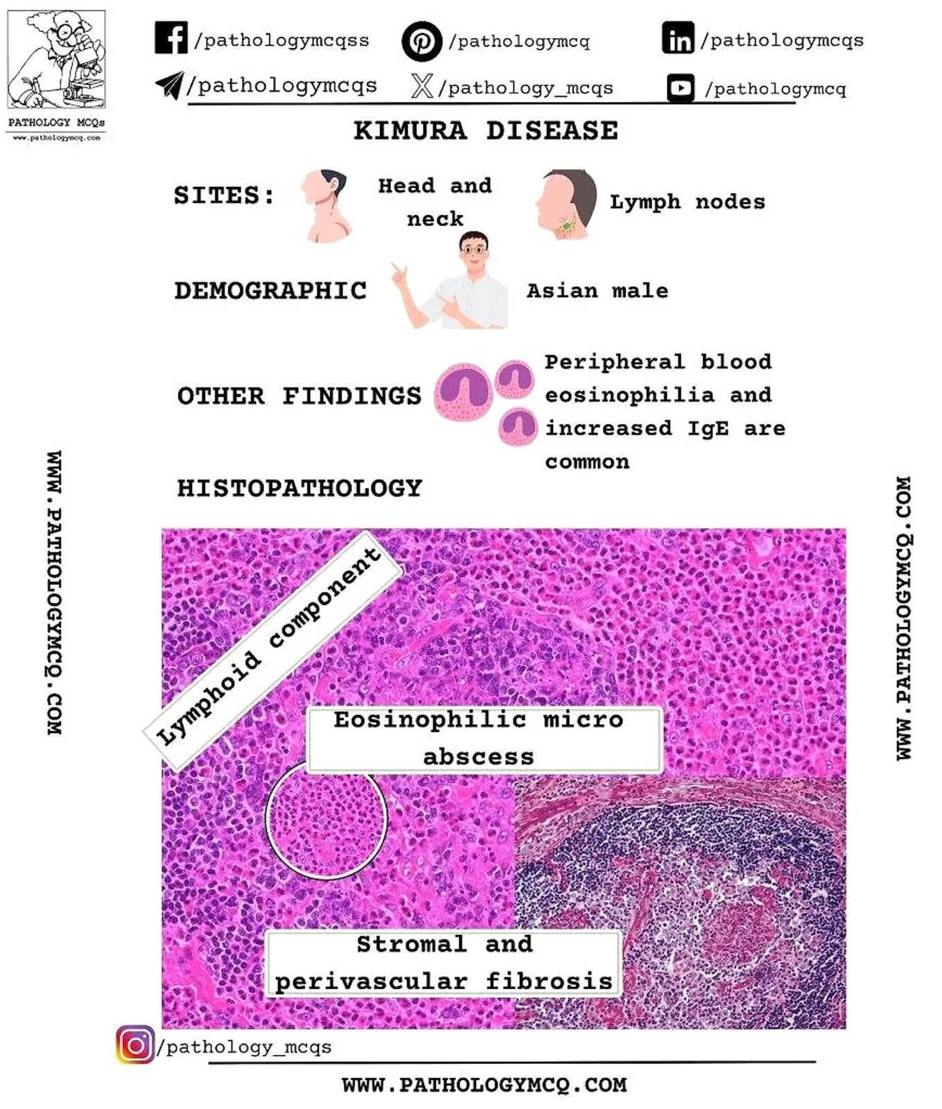
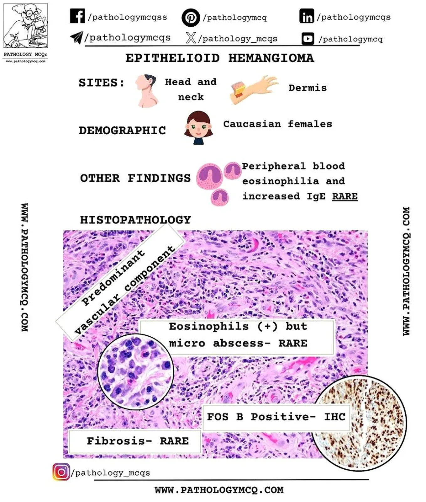
Kimura’s disease is a chronic inflammatory condition of unknown cause that affects young to middle-aged patients, most often males of Asian descent. Whereas, ALHE is more common in females.
2.LOCATION
Kimura disease patients usually have a mass in the head and neck region with involvement of subcutaneous tissue, soft tissue, salivary glands, and single or multiple regional lymph nodes. Epithelioid Hemangioma also commonly involves the head and neck region, particularly behind the ears.
3.HISTOLOGY
Key histologic features of kimura disease includes florid follicular hyperplasia that may contain a proteinaceous precipitate (IgE in a follicular dendritic network pattern) and vascularization of the germinal centers. The interfollicular areas show prominent high endothelial venules with a mixture of lymphocytes, plasma cells, eosinophils, and mast cells. Follicle lysis is often present, and eosinophilic abscesses are characteristic within germinal centers as well as in the paracortex.
Epithelioid Hemangioma on the other hand, is a vascular neoplasm characterized by the proliferation of blood vessels lined by plump endothelial cells with abundant eosinophilic cytoplasm, imparting a hobnail appearance. This lesion is part of the spectrum of what have been called histiocytoid or epithelioid hemangiomas, and is a low-grade vascular tumor. There is a dense, mixed inflammatory cell infiltrate consisting of lymphocytes, plasma cells, and eosinophils.
4. IMMUNOHISTOCHEMISTRY
Reticular pattern of IgE is seen in KIMURA disease. However, immunohistochemistry in Epithelioid Hemangioma is positive for CD 31, CD 34 and Factor VIII in the vascular component.
FOSB- IHC is usually positive in Epithelioid hemangioma
5.PERIPHERAL BLOOD EOSINOPHILIA
Peripheral blood examination shows eosinophilia and increased serum IgE levels in kimura disease but not so much in Epithelioid Hemangioma.
FIND A QUICK SUMMARY BELOW
https://www.pinterest.com/pin/1140114461886846593/sent/?invite_code=813193369a4b4d22b76b0347630622cd&sender=1140114599324959776&sfo=1REVIEW QUESTION
1. A 20- year- old Asian male has eosinophilia and high levels of IgE with cervical lymphadenopathy. Excisional biopsy of the lymph node demonstrates follicular hyperplasia, intense eosinophilia and eosinophilic microabscesses. h e most likely diagnosis is:
A. Kimura disease
B. Kikuchi- Fujimoto disease
C. Eosinophilic leukemia
D. Hypereosinophilic syndrome
CLICK TO REVEAL ANSWER
Answer is A- Kimura disease like Kikuchi disease is seen more ot en in the Asian population. h e etiology is not known. h is is a chronic inl ammatory disorder of the subcutaneous tissue and af ects regional lymph nodes. h e cervical area is the most common site to be involved. Histology of the af ected lymph nodes demonstrates follicular hyperplasia, eosinophilia with eosinophilic microabscesses and ini ltration of the germinal centers. Increase in vessels may also be seen. Warthin Finkeldey giant cells may also be present.
2. All are true about kimura disease except
A. Affects asians
B. Affects females
C. Peripheral eosinophilia is present
D. Elevated levels of IgE are seen
CLICK HERE TO REVEAL ANSWER
Answer is B- Kimura disease is more common in males.
Check out our hematopathology course and Hard copy notes for more updates and useful topics