
When it comes to diagnosing gastrointestinal diseases, Intestinal Tuberculosis (ITB) and Crohn’s Disease (CD) can pose a significant challenge due to their overlapping clinical and radiological features especially in south-east asian countries. However, understanding their macroscopic and microscopic differences can greatly aid in distinguishing between these two conditions.
Clinical Challenge: Why Differentiate?
Both ITB and CD present with similar symptoms such as abdominal pain, diarrhea, weight loss, and fever. Misdiagnosing one as the other can lead to inappropriate treatment, as ITB requires anti-tubercular therapy while CD is treated with immunosuppressants. Accurate diagnosis is, therefore, crucial to ensure the best patient outcomes.
Macroscopic Features
Intestinal Tuberculosis:
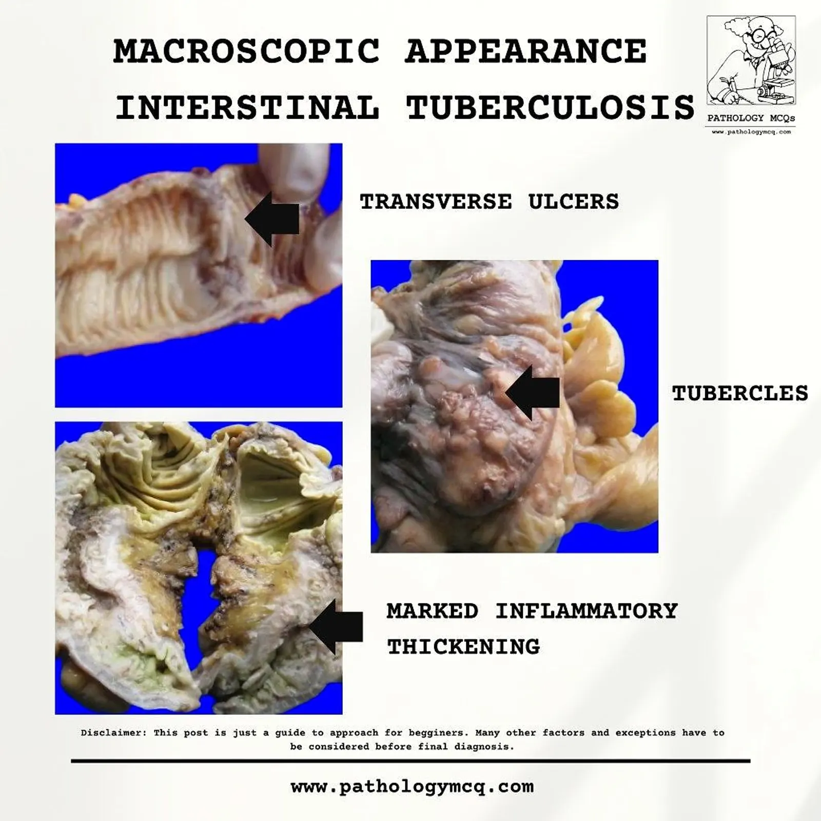
- Transverse Ulcers: Ulcers in ITB are characteristically transverse, involving the circumference of the bowel.
- Tubercles: Visible nodular lesions caused by granulomatous inflammation.
- Marked Inflammatory Thickening: The bowel wall is often thickened with significant fibrosis, causing stricture formation.
Crohn’s Disease:
- Cobblestone Mucosa: A hallmark feature due to ulceration and swelling of the intestinal mucosa.
- Creeping Fat: Mesenteric fat wraps around the bowel, often extending to the serosa.
- Deep Fissuring Ulcers: Linear and deep ulcers that penetrate the bowel wall.

Microscopic Features
Intestinal Tuberculosis:
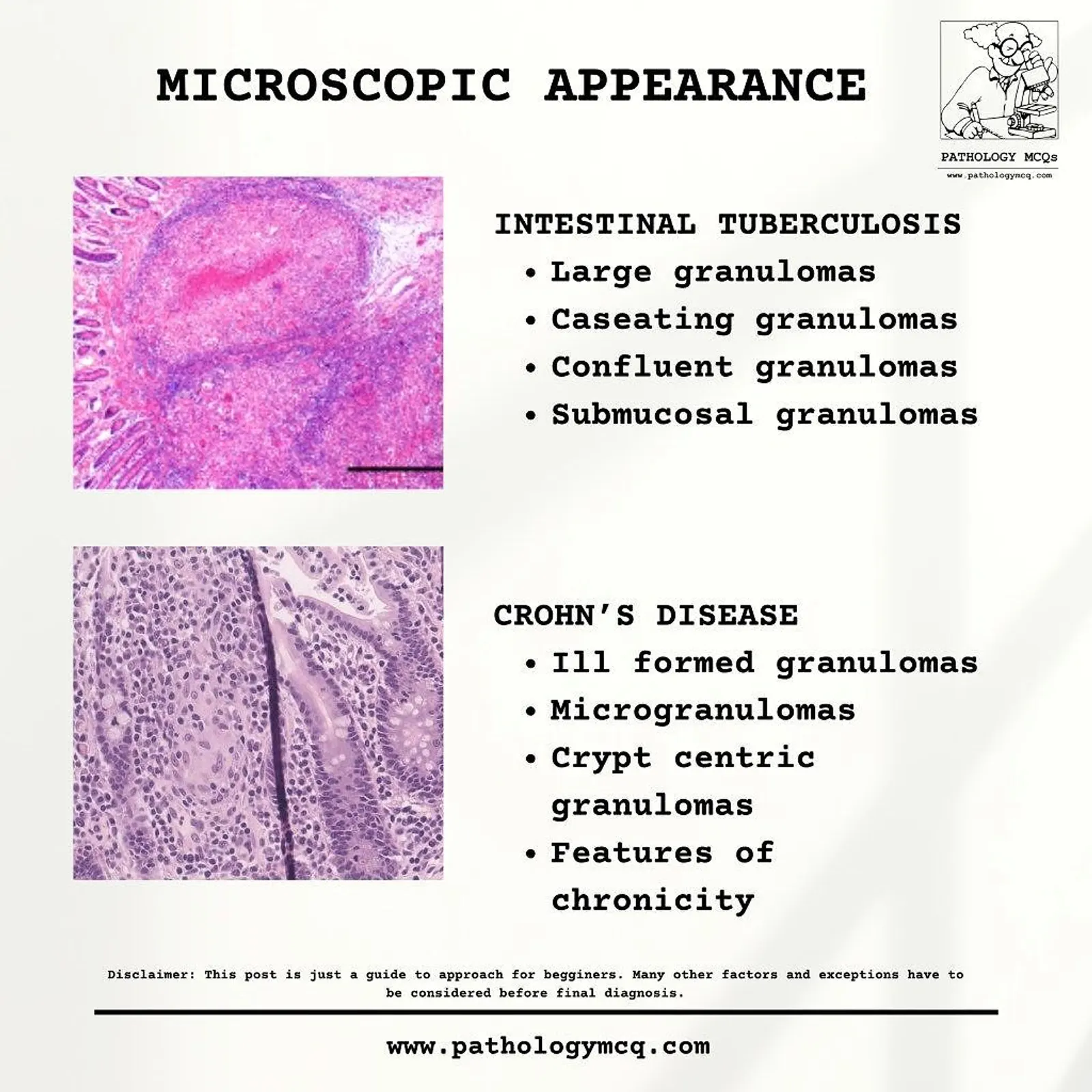
- Granulomas: Large, confluent, and caseating granulomas, often extending to the submucosa and beyond.
- Epithelioid Cells: Granulomas in ITB are composed of epithelioid cells surrounded by lymphocytes.
- Acid-Fast Bacilli (AFB): Staining (e.g., Ziehl-Neelsen) reveals Mycobacterium tuberculosis.
Crohn’s Disease:
- Ill-Formed Granulomas: Non-caseating granulomas are smaller and scattered.
- Crypt-Centric Granulomas: Granulomas often surround crypts.
- Transmural Inflammation: CD shows inflammation throughout the bowel wall layers, unlike the localized granulomas in ITB.
Key Diagnostic Features:
| Feature | Intestinal Tuberculosis | Crohn’s Disease |
|---|---|---|
| Granulomas | Caseating, large, confluent | Non-caseating, ill-formed, scattered |
| Ulcers | Transverse | Linear, deep fissuring |
| Involvement | Localized, common in ileocecal region | Segmental, "skip lesions" |
| Fibrosis | Marked | Less prominent |
| Mesenteric Fat Involvement | Absent | Creeping fat |
| Ziehl-Neelsen Staining | Positive (AFB present) | Negative |
Approach to Diagnosis
- Clinical History and Imaging: While imaging may suggest either ITB or CD, definitive diagnosis relies on pathology.
- Biopsy and Histopathology: Tissue biopsy is essential for identifying the granulomatous inflammation characteristic of both diseases.
- Microbiological Studies: AFB staining and PCR for Mycobacterium tuberculosis are key in confirming ITB.
- Response to Treatment: In challenging cases, a therapeutic trial with anti-tubercular drugs may help distinguish ITB from CD.
Conclusion
Differentiating between Intestinal Tuberculosis and Crohn’s Disease is critical for effective management. While there are significant overlaps, recognizing the differences in macroscopic and microscopic appearances can guide pathologists and clinicians toward an accurate diagnosis. Always correlate histopathological findings with clinical and microbiological data for a comprehensive evaluation.
Disclaimer: This blog is intended as an educational resource for medical students, pathology residents and professionals. Always consider multiple factors before arriving at a final diagnosis.
Video explanation
FOR MORE POINTS LIKE THESE
Join our approach based course