
Subtitle: A complete high-yield review of apoptosis with exam-focused MCQs, images, and takeaways
Author: Pathology MCQ Editorial Team
Category: General Pathology
Read Time: 7–10 minutes
At a glance
- Mechanisms of apoptosis: intrinsic, extrinsic, mitochondrial pathways
- Key regulators: Bcl-2 family, caspases, death receptors
- Classic microscopic features with images
- 15 high-yield MCQs with simple explanations
Contents
- Introduction
- Mechanisms of Apoptosis
- Morphologic Features of Apoptosis
- Physiologic & Pathologic Apoptosis
- 10 Robbins-Level MCQs
- Key Takeaways
- Recommended Learning
- References
1. Introduction
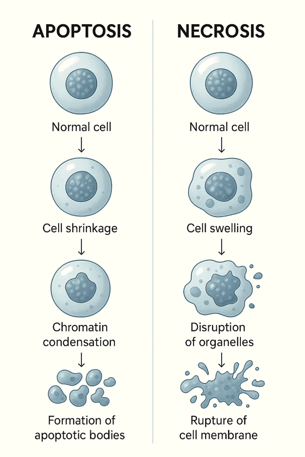
Apoptosis is a controlled, ATP-dependent programmed cell death mechanism essential for maintaining tissue homeostasis. Unlike necrosis, it is non-inflammatory and characterized by cell shrinkage, chromatin condensation, and formation of apoptotic bodies. Failure of apoptosis contributes to cancer, autoimmune disease, and degenerative disorders.
2. Mechanisms of Apoptosis
A. Intrinsic (Mitochondrial) Pathway
Triggered by:
- DNA damage
- ER stress
- Hypoxia
- Growth factor withdrawal
Regulated by Bcl-2 family proteins:
- Anti-apoptotic: Bcl-2, Bcl-XL, Mcl-1
- Pro-apoptotic: Bax, Bak
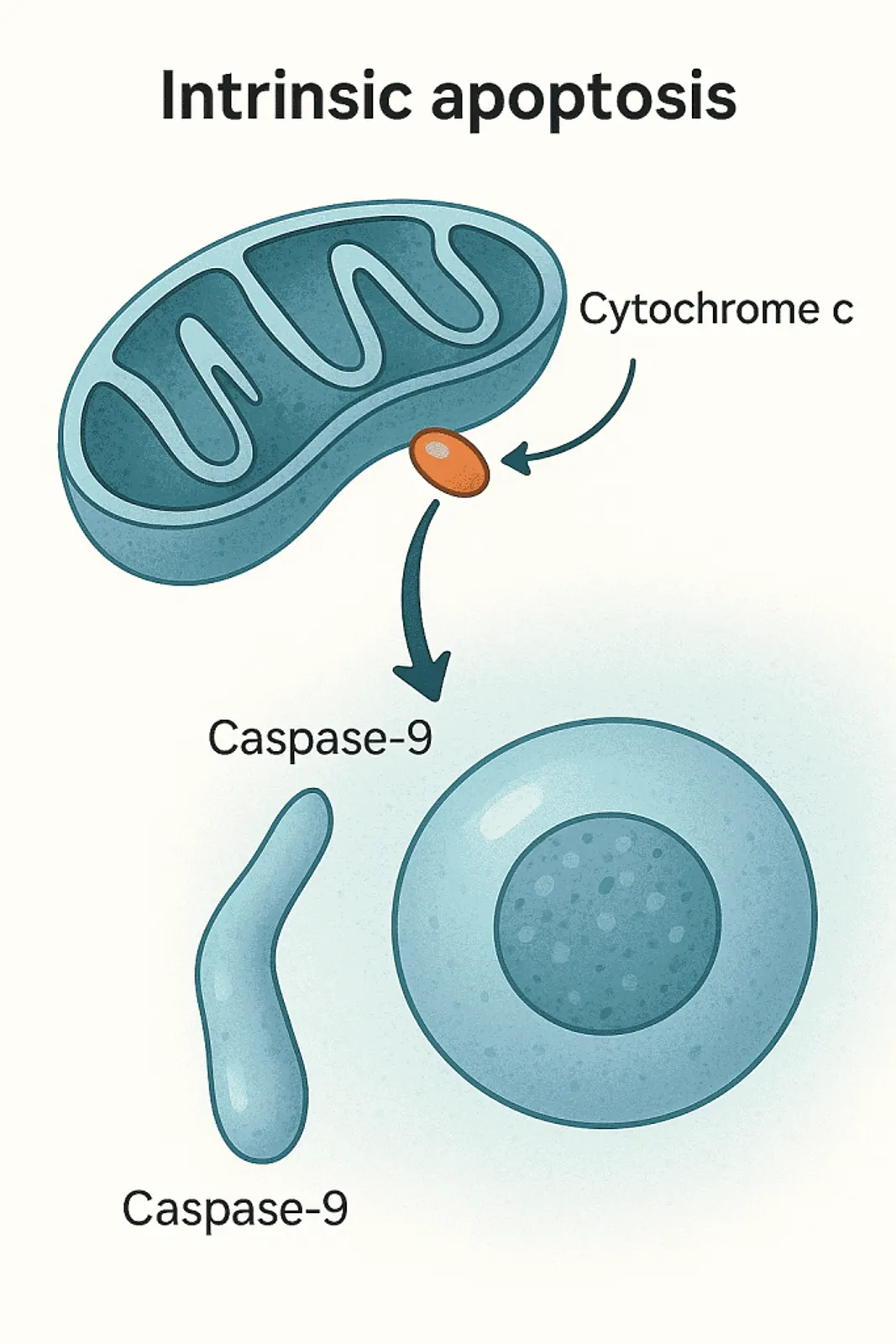
Release of cytochrome c activates caspase-9, forming the apoptosome.
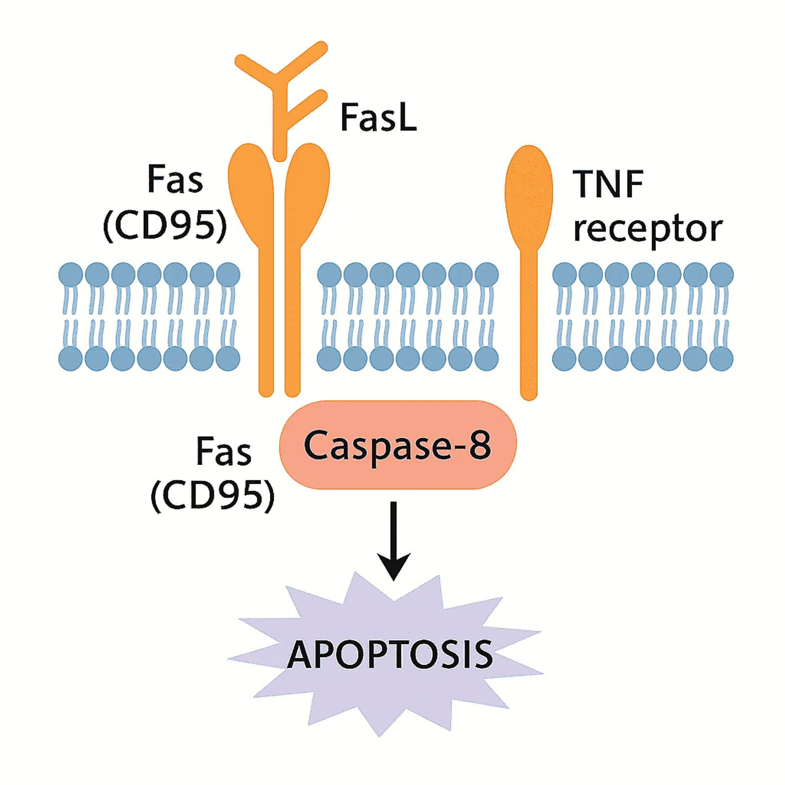
B. Extrinsic (Death Receptor) Pathway
Initiated via:
- Fas (CD95)
- TNF receptor
Binding of FasL or TNF leads to activation of caspase-8.
Important in:
- Immune elimination of infected cells
- Tumor surveillance
C. Execution Phase
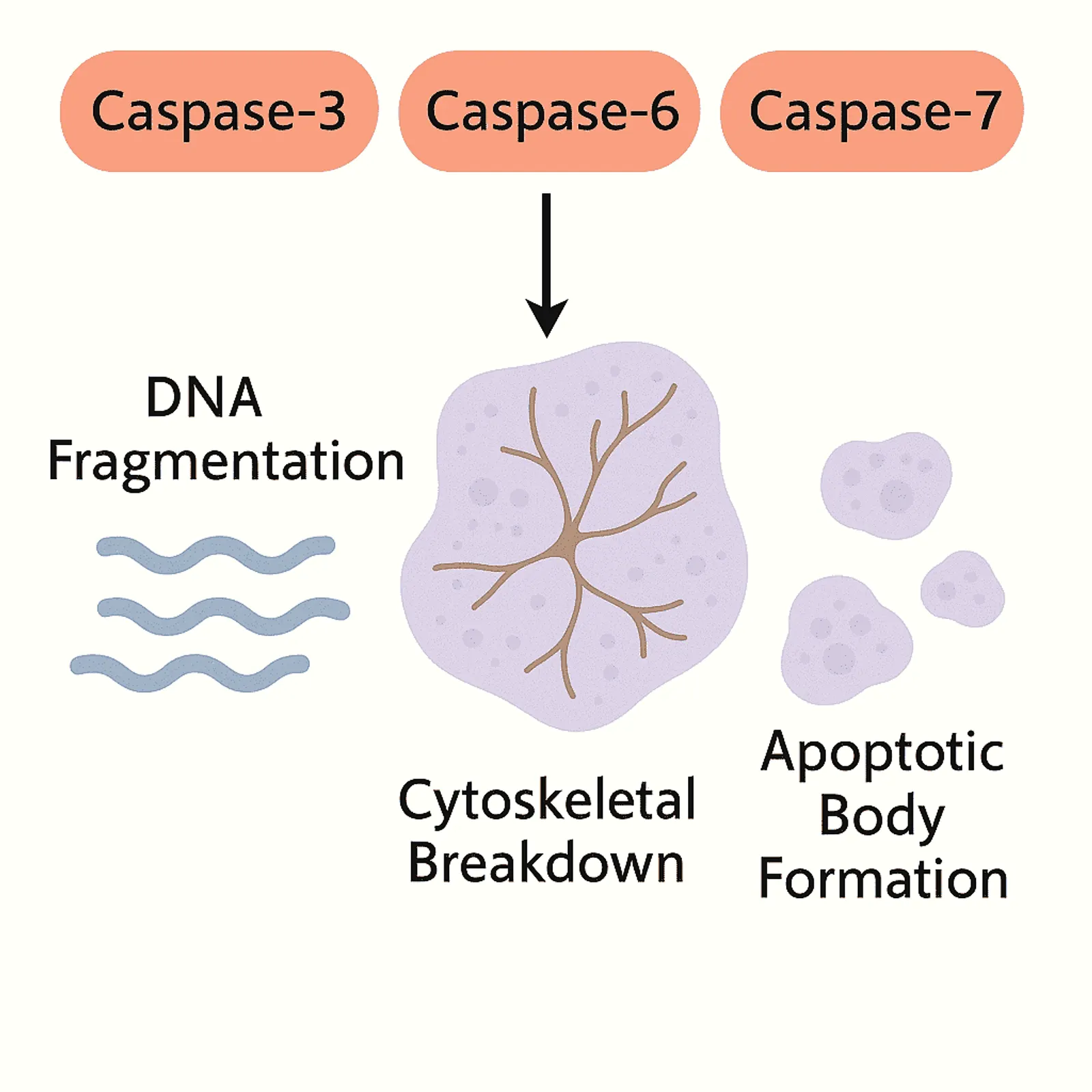
Common downstream pathway:
- Activation of caspase-3, 6, 7
- DNA fragmentation
- Cytoskeletal breakdown
- Apoptotic body formation
3. Morphologic Features of Apoptosis
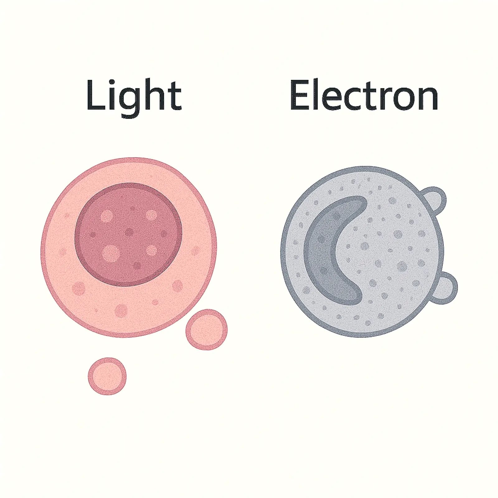
Light Microscopy
- Cell shrinkage
- Deep eosinophilia
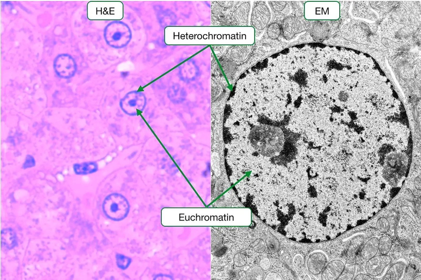
- Chromatin condensation (pyknosis, karyorrhexis)
- Rounded eosinophilic apoptotic bodies
- No inflammation
Electron Microscopy
- Chromatin crescent formation
- Intact cell membrane
- Membrane blebs
4. Physiologic & Pathologic Apoptosis
Physiologic
- Embryogenesis (digit separation)
- Hormone withdrawal (endometrium, breast)
- Negative selection of T-cells
- Involution of thymus
Pathologic
- DNA damage (radiation, chemotherapy)
- Viral infections
- Misfolded protein accumulation (ER stress)
- Cell death in neurodegenerative diseases
5. 10 Robbins-Level MCQs
[mlw_quizmaster quiz=495]6.Key takeaways
- Intrinsic pathway uses caspase-9, extrinsic uses caspase-8
- Bcl-2 inhibits apoptosis; Bax/Bak promote it
- Morphology: cell shrinkage, apoptotic bodies, no inflammation
- Apoptosis is ATP-dependent and orderly
7. Recommended learning
8. References
- Robbins & Cotran Pathologic Basis of Disease
- WHO Classification of Tumours (IARC)
- Kumar, Abbas, Aster – Basic Pathology
- NEJM Review on Cell Death Pathways