
Subtitle: Detailed review of hepatocellular carcinoma subtypes: WHO classification, prognosis table, molecular markers, histology. Pathology exam prep with MCQs
Author: Pathology MCQ Editorial Team
Category: Hepatopathology & Oncopathology
Last Updated: March 2026
Read Time: 18 min
This is a medical, educational, exam-oriented pathology review focused on diagnosis and histopathology of hepatocellular carcinoma subtypes.
At-a-Glance
- 8 WHO subtypes: Steatohepatitic (freq 6-36%), clear cell (better prog), MTM (worse), etc..
- Key exam link: Fibrolamellar DNAJB1-PRKACA fusion; MTM TP53.
- Prognostic spectrum: Lymphocyte-rich best; neutrophil-rich worst.
Difficulty Level: Moderate to Difficult
Table of Contents
Overview of Hepatocellular Carcinoma Subtypes
Hepatocellular carcinoma subtypes constitute morphologically defined variants recognized in the WHO Classification of Tumours: Digestive System Tumours (5th edition, 2019), comprising approximately 35% of cases. These include steatohepatitic, clear cell, macrotrabecular-massive (MTM), scirrhous, chromophobe, fibrolamellar, neutrophil-rich, and lymphocyte-rich HCC histological variants. Prognosis varies significantly: MTM and neutrophil-rich portend poor outcomes, while clear cell and lymphocyte-rich fare better. Accurate subtyping via histology, IHC, and molecular profiling refines staging and therapy.
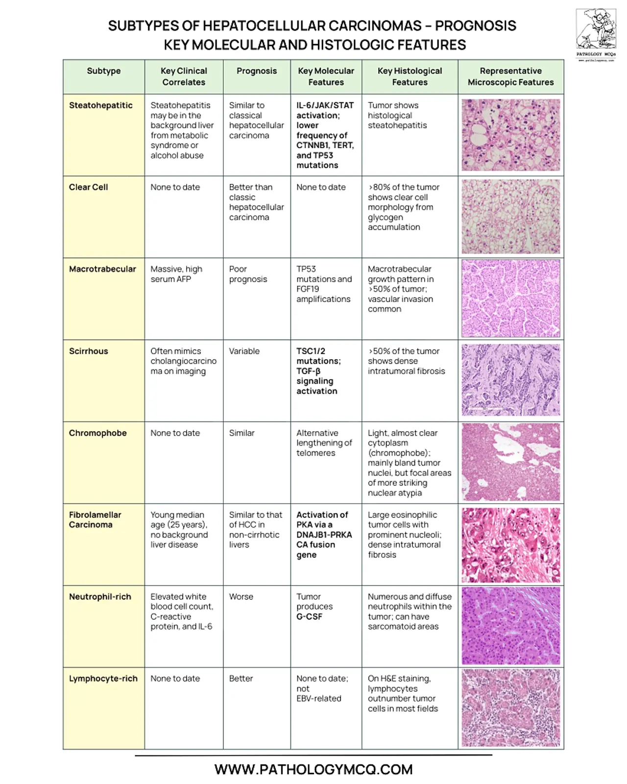
This image meticulously tabulates HCC subtypes: steatohepatitic (steatosis freq 6-36%, similar prog, steatohepatitis-like); clear cell (no abuse, better prog, 80% tumor glycogen); macrotrabecular massive (AFP high, poor, TP53/mut amp); scirrhous (often imaging var, similar, TGFb1/12 stroma); chromophobe (none to date, similar, light clear focal atypia); fibrolamellar (young no cirr, similar, DNAJB1-PRKACA CA); neutrophil-rich (elev WBC, worse, G-CSF num diff); lymphocyte-rich (no to date, better, CD8+ stroma). Diagnostic value: Instant prognostic correlation; essential for pathology board exams; highlights stains like TFE3 implied for chromophobe.
WHO Classification Framework
WHO mandates subtyping when a pattern dominates ≥50% tumor volume, integrating Robbins-described features like architectural atypia. Genomic underpinnings: Steatohepatitic low CTNNB1; MTM high TP53 (50-70%). Frequency data from large series: Steatohepatitic most common variant.
Steatohepatitic HCC
Steatohepatitic HCC requires ≥5% tumor with steatosis, hepatocyte ballooning, Mallory-Denk hyaline, lobular inflammation, and pericellular fibrosis. Frequency 6-36%; prognosis equivalent to conventional HCC (5-yr survival ~30%). Molecular: IL6/JAK/STAT pathway activation, TP53/CTNNB1 mutations rare (<20%). Clinical: NAFLD/obesity association; histology mimics background liver disease.
Clear Cell HCC
Clear cell HCC defined by ≥80% tumor cells with optically clear cytoplasm due to glycogen accumulation. Prognosis superior (better survival post-resection); frequency 3-7%. Molecular hallmarks: IDH1/2 mutations (3-6%), low TERT promoter alterations. IHC retains hepatocellular markers (HepPar-1, arginase-1); pitfalls include renal cell mimicry.
Macrotrabecular-Massive HCC
MTM-HCC characterized by ≥50% tumor with trabecular architecture ≥10 cells thick. Prognosis poorest among subtypes (HR 2.5 for recurrence); frequency ~5%. Molecular: TP53 mutations (frequent), MYC amplification, high AFP (>100 ng/mL). Associations: HBV, advanced AJCC stage.
Scirrhous HCC
Scirrhous HCC features dense fibrous stroma occupying >50% tumor volume with embedded thin cords/sinusoids. Prognosis similar to conventional; frequency ~4%. Molecular: TGF-β signaling, cholangiocyte markers (CK7/19+), stem cell features. Histology: Often infiltrative, mimicking intrahepatic cholangiocarcinoma.
Chromophobe HCC
Chromophobe HCC displays uniform chromophobic (pale) cytoplasm with minimal pleomorphism and focal atypia. Prognosis akin to conventional; frequency ~3%. Molecular data limited; possible TFE3 overexpression. Rare; requires excluding oncocytic variants.
Fibrolamellar HCC
Fibrolamellar HCC presents in young (<30y) without cirrhosis, with large polygonal eosinophilic cells intersected by thick lamellar fibrosis. Prognosis similar overall but favorable if resectable (5-yr 70%); frequency 1-5%. Pathognomonic: DNAJB1-PRKACA gene fusion (>90%). IHC: CK7+, pale bodies common.
Neutrophil-rich & Lymphocyte-rich HCC
Neutrophil-rich HCC: Prominent neutrophilic infiltrate (no abscess), elevated WBC/CRP; worse prognosis. Possible G-CSF production. Lymphocyte-rich HCC: Dense CD8+ lymphocytic stroma (>50%); better prognosis, immunotherapy responsive.
Comprehensive Prognosis Table
Molecular Diagnostics
NGS panels detect fusions (FLC), mutations (TP53 MTM); IHC panels: Glypican-3, CD34 vascular. Prognostic: High Ki-67 in MTM.
Histopathologic Reporting
Report subtype if dominant; note % involvement, background liver. Synoptic: Pattern, IHC, molecular.
High - yield MCQS
[qsm quiz=592]Exam Pearl
Steato=similar/steatosis; Clear=better/glycogen; MTM=poor/trabeculae; FLC=fusion/young.
Key Takeaway
Subtyping HCC per WHO prognosticates: MTM adverse, clear cell favorable—integrate histology/molecular for reports.