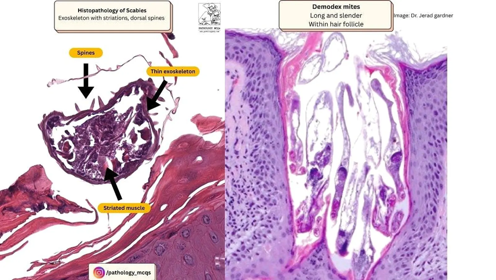
Scabies is a parasitic infestation caused by Sarcoptes scabiei, a mite that burrows into the epidermis, leading to intense pruritus and inflammation. Diagnosis can be clinched with a keen eye on histopathological hallmarks. This blog explores the diagnostic features of scabies under the microscope using detailed, labeled histology images and a whole slide image viewer for interactive exploration.
What Does Scabies Look Like on Histology?
Below are the key features you should look for while diagnosing scabies histologically:
1. Mites in the Stratum Corneum
- Sarcoptes scabiei can be seen as oval, refractile bodies burrowing within the stratum corneum.
- These mites measure approximately 0.2–0.4 mm in length.
- Their presence in the superficial layers of skin is a hallmark finding.

2. Mite Morphology: A Closer Look
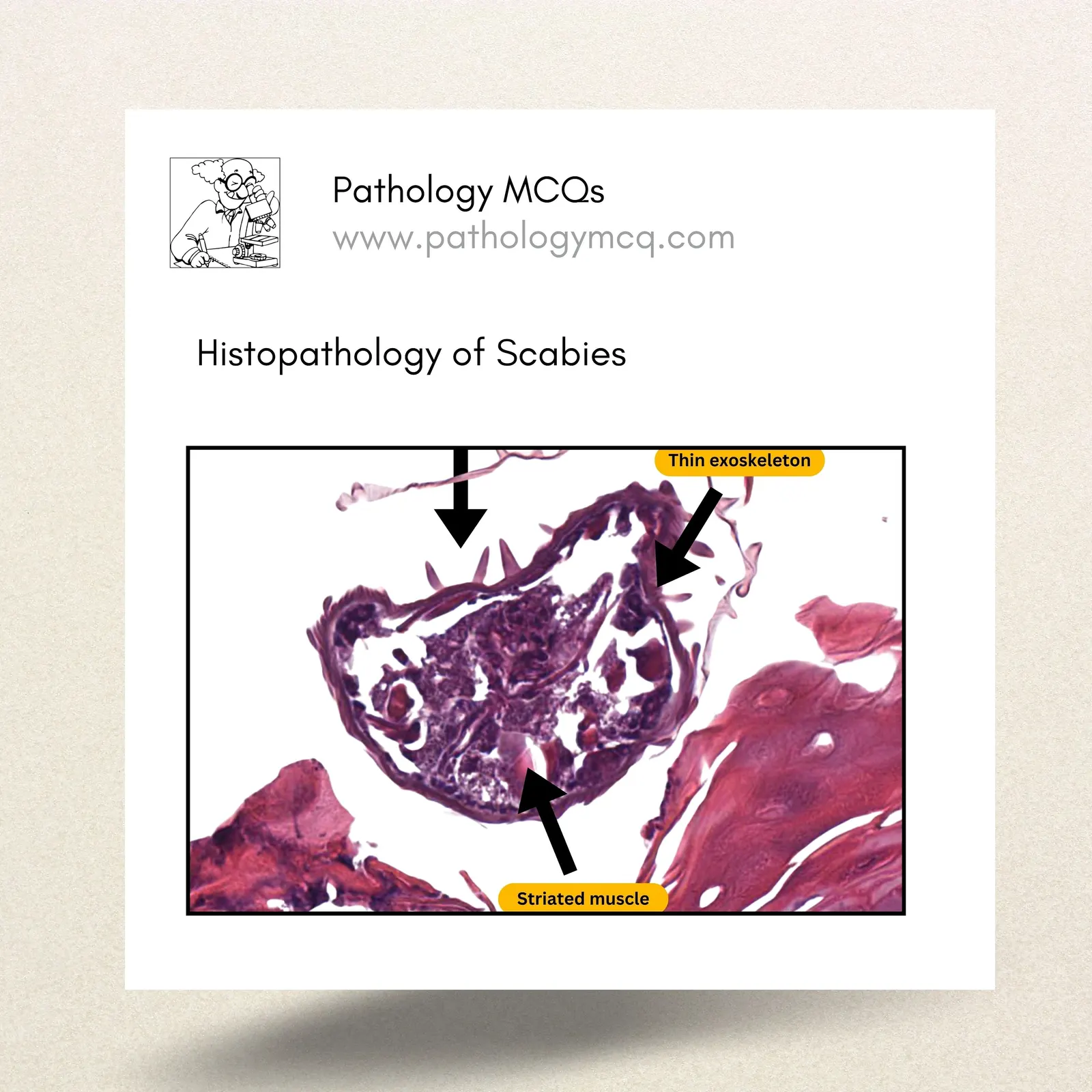
- On higher magnification, the mite exhibits:
- A thin exoskeleton
- Internal striated muscle fibers
- External dorsal spines, a distinctive morphological clue
- Additionally, 4 legs can also be identified.

3. Egg Structures
- Scabies eggs are oval and measure 0.1–0.2 mm.
- They can be found within the stratum corneum, often with a visible egg shell.

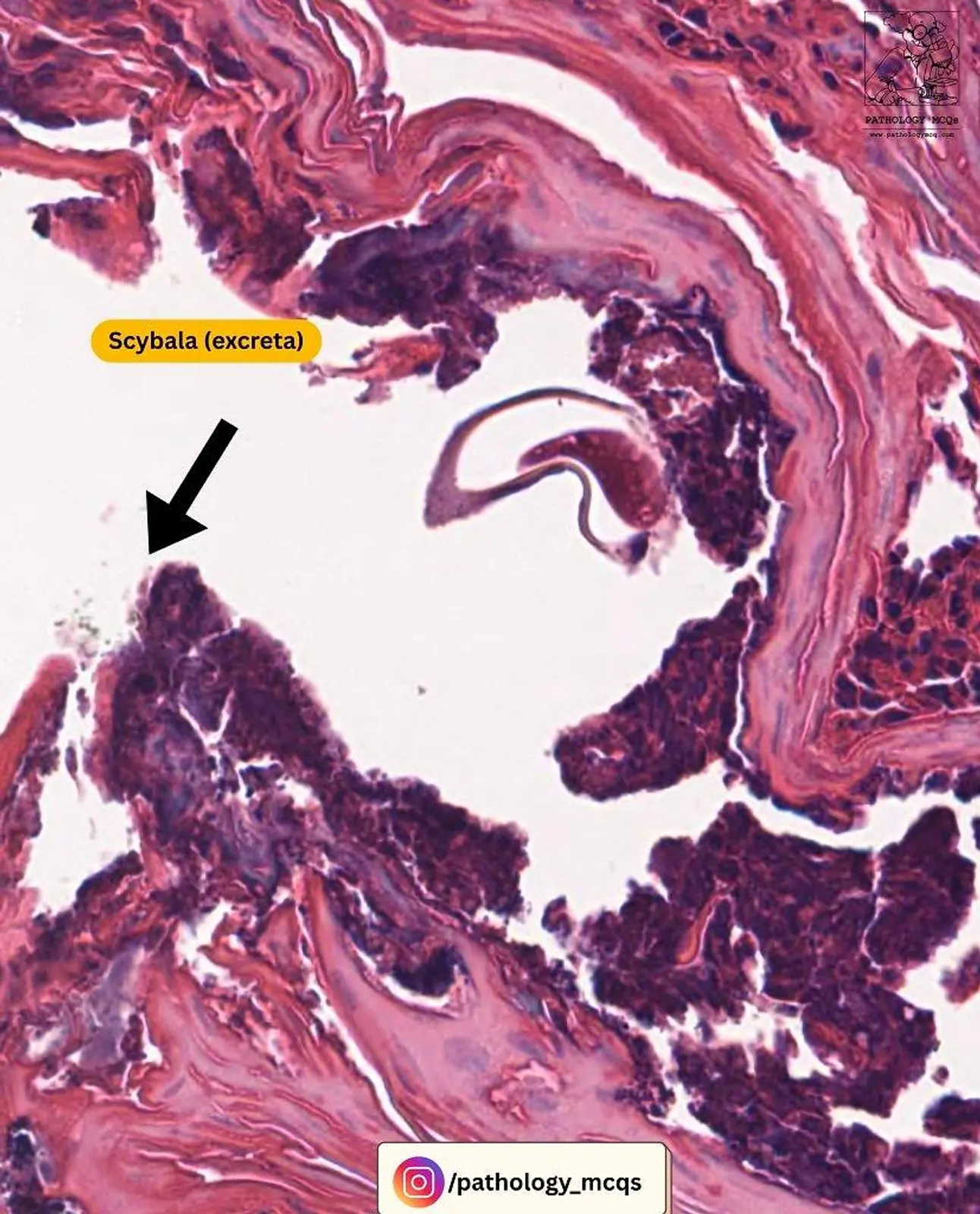
4. Scybala: Mite Droppings
- Scybala (fecal pellets) appear as dark granular clumps in the burrow and incite an inflammatory reaction.
5. Inflammatory Reaction
- The host immune response is characterized by:
- Neutrophilic microabscesses around the mite
- Perivascular lymphoeosinophilic infiltrate with scattered histiocytes

Explore the Case on Whole Slide Viewer
You can interact with the entire scanned slide using the viewer below. Zoom into the stratum corneum to trace the mite’s path, inspect the burrows, or look for eggs and scybala.
QUICK TIPS
- Always scan the stratum corneum carefully — mites never go deeper than that.
- Look for multiple clues: mite body, eggs, scybala, and inflammatory response.
- Use high-power fields to confirm exoskeletal spines and striated muscle in the mite.
Finally!! Some differences between - Scabies and Demodex mite.
| Feature | Scabies Mite (Sarcoptes scabiei) | Demodex Mite (Demodex folliculorum) |
| Location in Skin | Stratum corneum | Hair follicles, deeper folllicl and sebaceous glands |
| Size | 0.2 – 0.4 mm (short and broad) | ~0.3 – 0.4 mm (long and slender) |
| Body Shape | Round to oval, tortoise-like | Elongated, worm-like (cigar-shaped) |
| Legs | 4 pairs anteriorly | 4 pairs anteriorly (but less prominent) |
| Visible Internal Structures | Striated muscle, gut, spines | Less distinct internal detail |
| Associated Pathology | Burrows with inflammation, eggs, scybala | Folliculitis, rosacea-like inflammation |
| Staining Visibility | Well-visualized on H&E | Seen on H&E but better with oil-immersion prep |