
Historically, p53 IHC has been interpreted as negative or positive based on the percentage of stained tumor cell nuclei using variable cut-offs which range from 5 to 50%.
More recently, a tripartite interpretation has been suggested, wherein an “overexpressed or no expression (all or nothing)” nuclear staining pattern is highly predictive of underlying TP53 mutation while a normal/wild type pattern is not.
Of note, rare cases with TP53 mutation rarely show cytoplasmic staining, and, about 5% cases have a wild type pattern
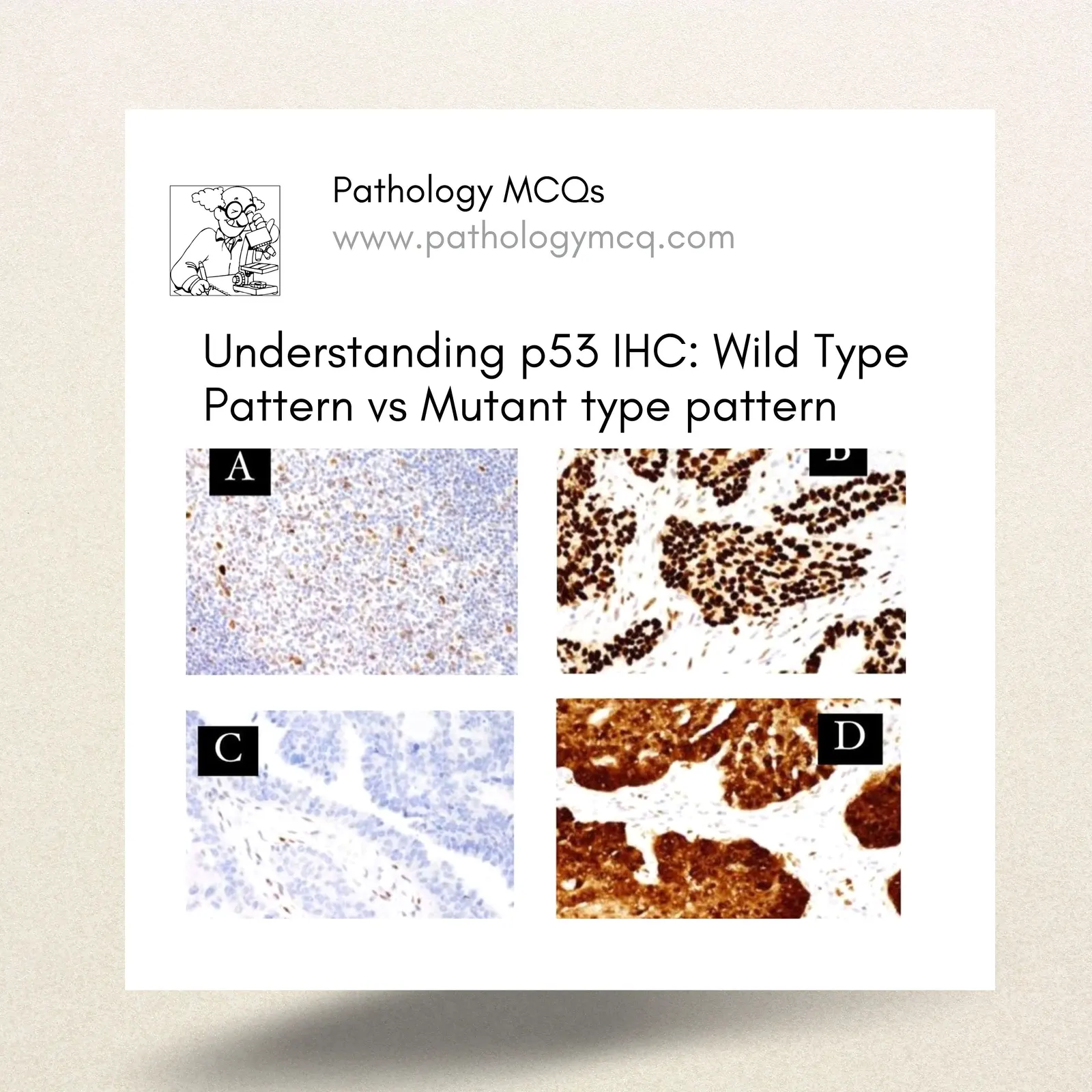
- WILD TYPE P53 (NORMAL)
The distribution of nuclear staining in a 'wild type' pattern ranges from a few positive cells to almost all cells staining, but with variable intensity.
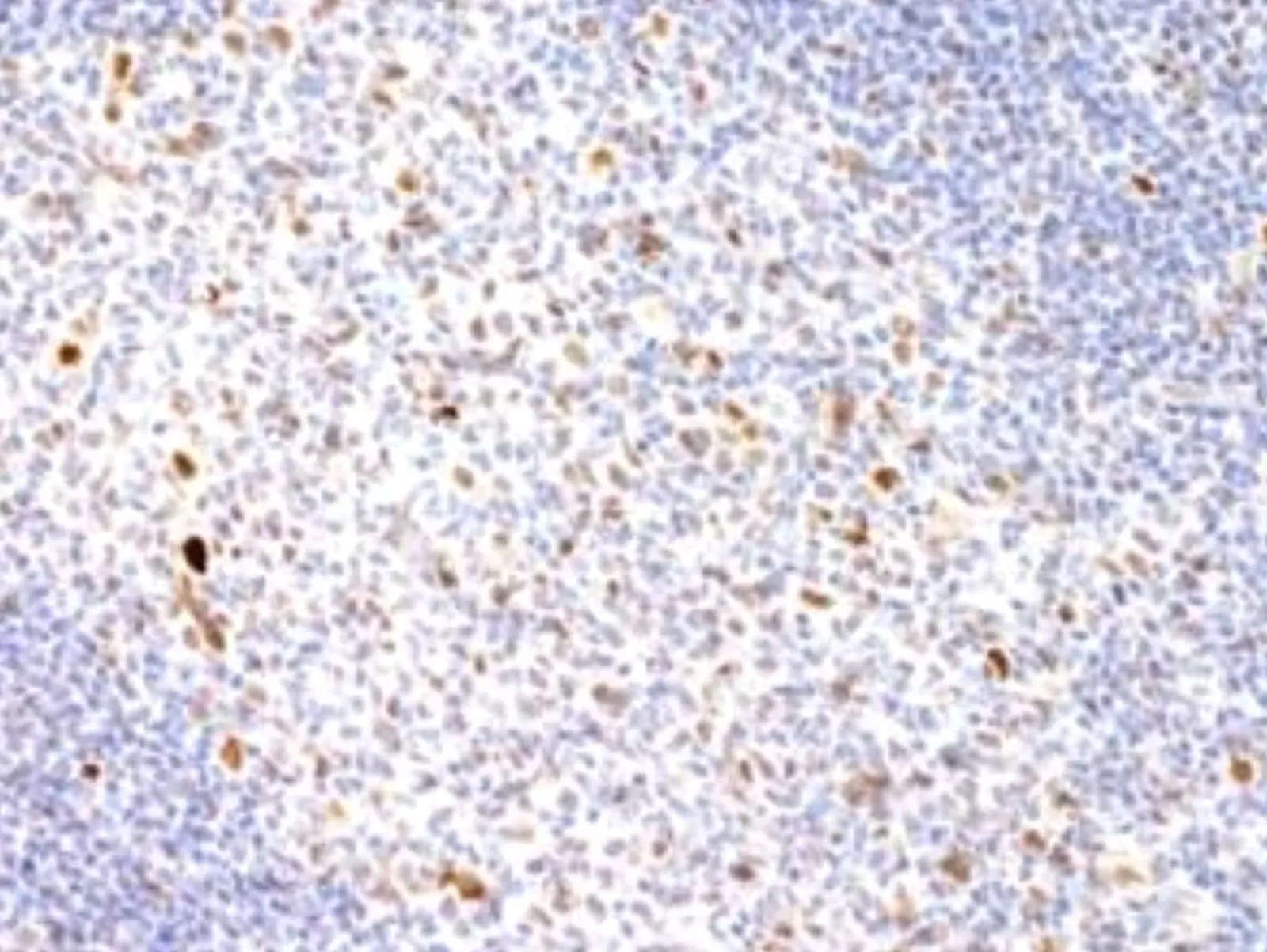
2. OVEREXPRESSION (ABNORMAL p53)
- Overexpression is defined as strong nuclear staining in at least 80% of tumor cell nuclei (usually 100% tumor cell nuclei, the 80% cut-off accounts for technical artifacts and the rare mosaic pattern, see below).
- Overexpression is the most common (~66%) pattern observed in HGSC, almost certainly signifying an underlying TP53 mutation.
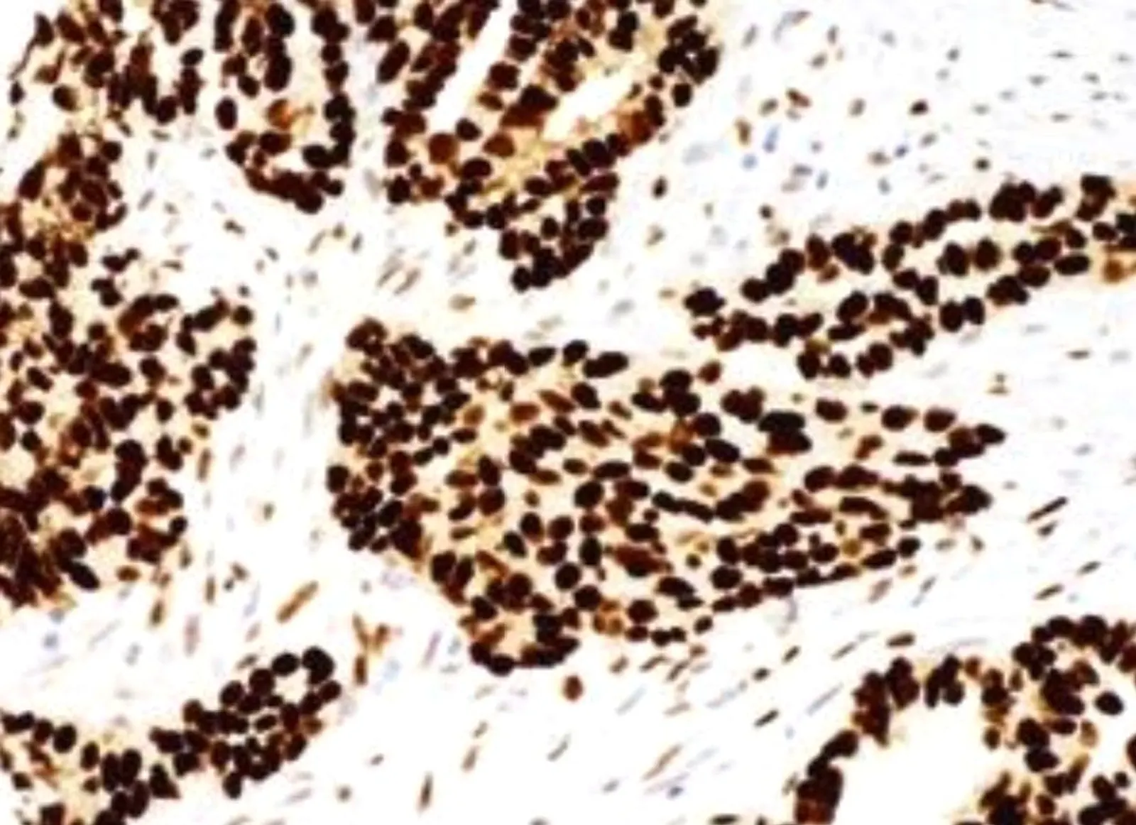
3. COMPLETE ABSENCE OR NULL PATTERN (ABNORMAL)
- Complete absence is defined as no staining in tumor cell nuclei in the presence of
'wild-type', ie variable, staining in normal background cells (intrinsic control present).
The interpretation is similar to that of DNA mismatch repair gene protein expression. - Complete absence is observed in ~25% of HGSC.
- Complete absence is almost certainly indicative of underlying TP53 mutation.
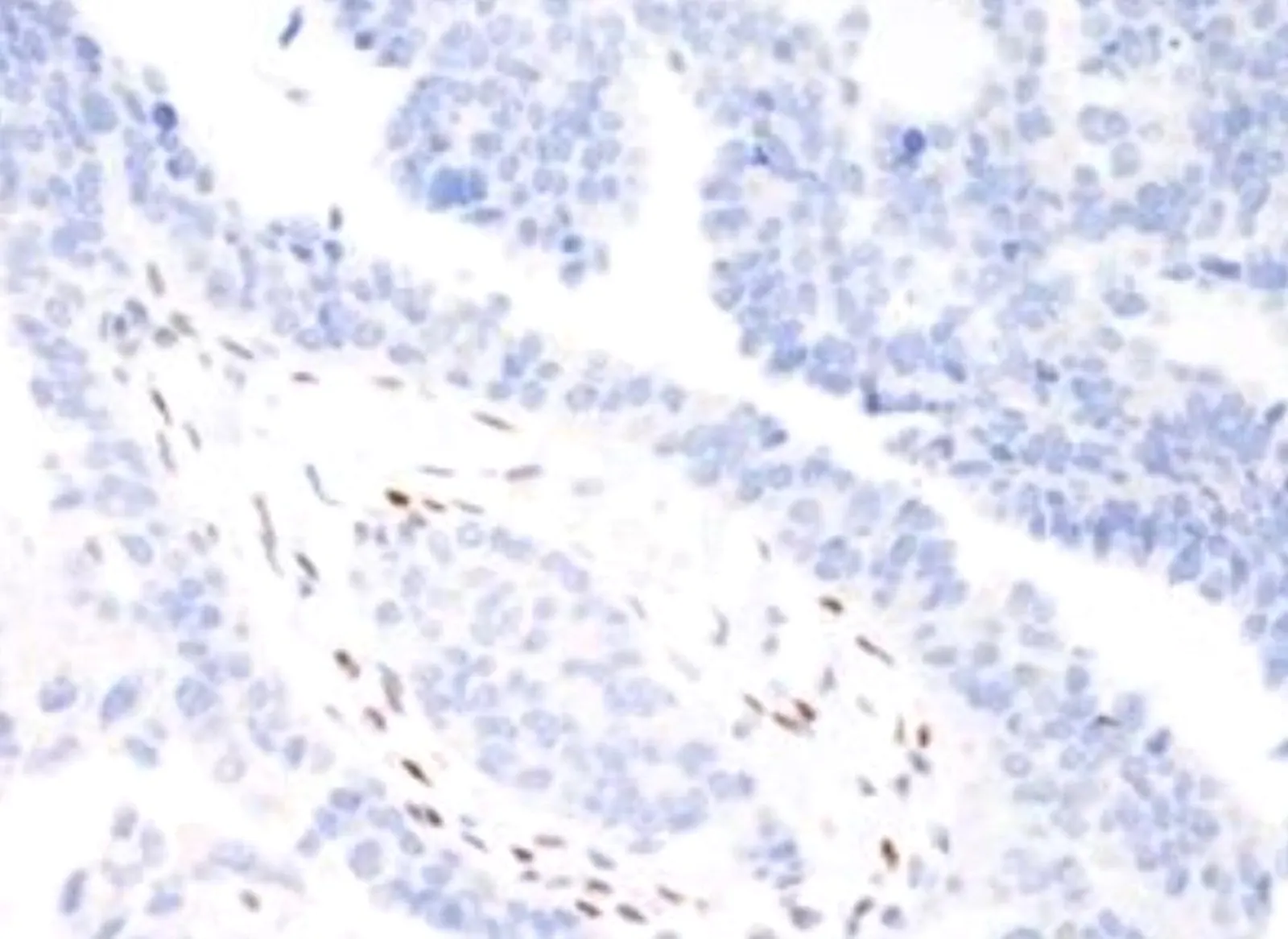
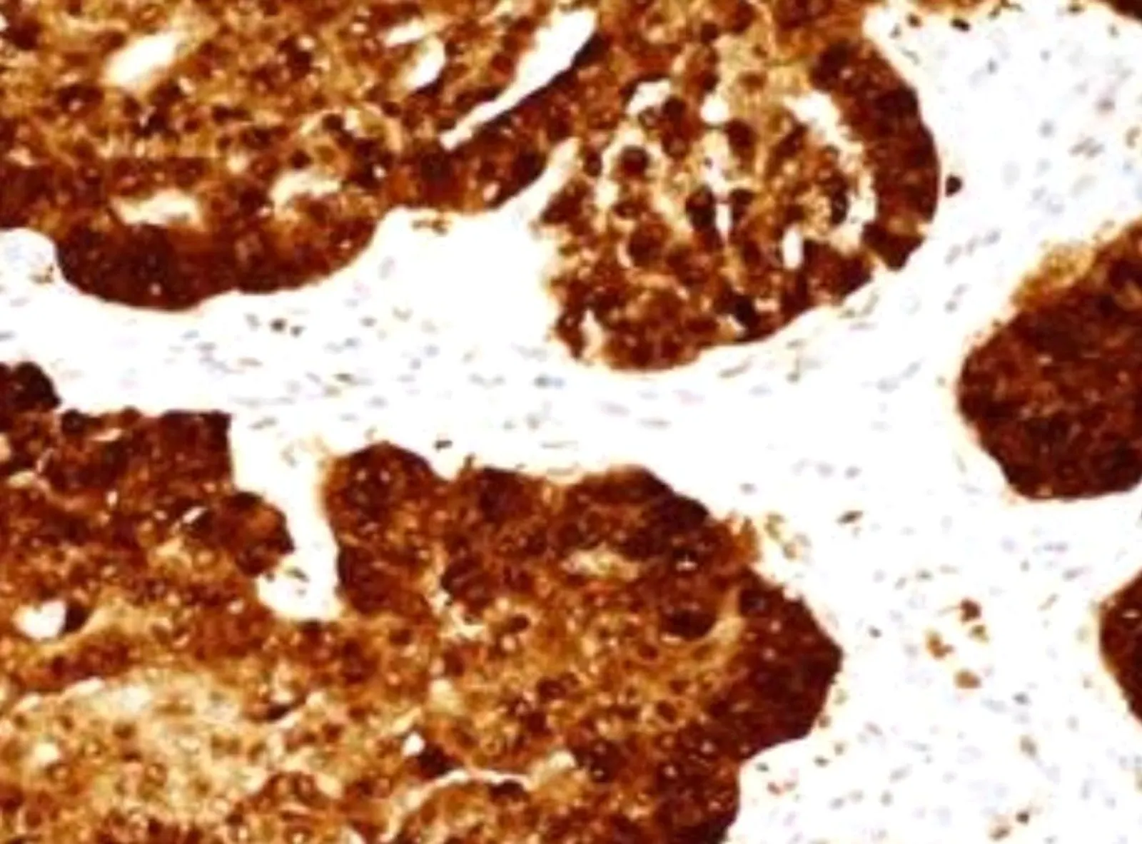
4. CYTOPLASMIC (ABNORMAL)
- Cytoplasmic staining is defined as predominant cytoplasmic staining in the absence of strong nuclear staining in >80% of tumor cell nuclei, i.e. the nuclear staining can be absent or weak and variable.
- The cytoplasmic pattern is seen in ~4% of high-grade serous carcinomas and is associated with underlying TP53 mutation.
Try to answer this MCQ now
Correct answer is : Option A -wild type p53. Very rare in high grade serous carcinomas, whereas wild type p53 is seen in low grade serous carcinomas.
FOR MORE DIAGNOSTIC INSIGHTS, JOIN US IN OUR COURSE
TO ACE YOUR FRCPath EXAMS AND CONQUERING MCQs WITH CRITICAL THINKING, JOIN US IN OUR FRCPath COURSE