
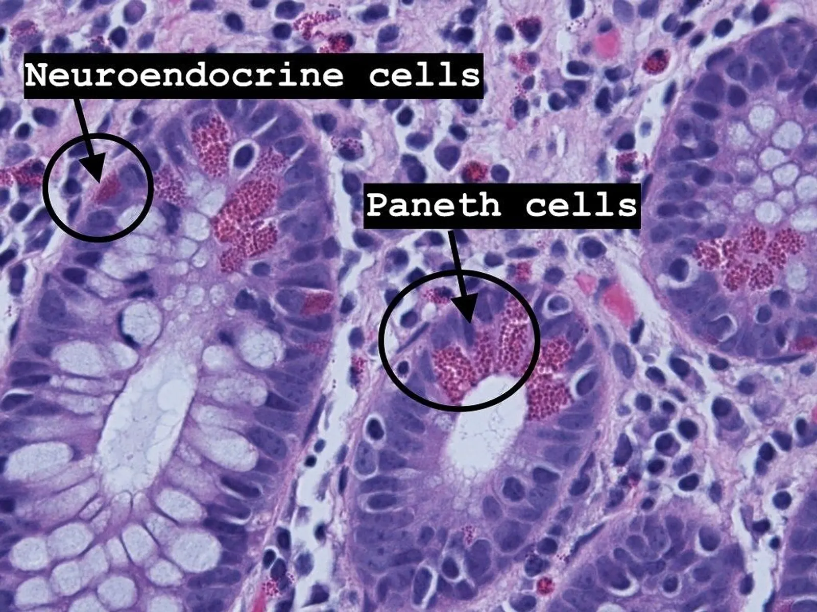
Understanding the histological features and clinical significance of Paneth cells and neuroendocrine cells is crucial for pathologists interpreting colonic biopsies. This blog delves into their distribution, morphology, and roles, highlighting their importance in diagnosing intestinal diseases.
TRY TO ANSWER THESE MCQS BEFORE MOVING ON
What are Paneth Cells and Neuroendocrine Cells?
Paneth Cells
- Specialized epithelial cells located at the base of crypts.
- Predominantly found in the small intestine but can appear in the colon in pathological conditions.
- Contain eosinophilic granules rich in antimicrobial peptides like lysozyme.
Neuroendocrine Cells
- Part of the diffuse neuroendocrine system scattered throughout the gastrointestinal tract.
- Secrete hormones like serotonin, somatostatin, and VIP to regulate intestinal physiology.
- Require immunohistochemical markers (e.g., chromogranin A, synaptophysin) for identification.
Key Differences Between Paneth Cells and Neuroendocrine Cells
| Feature | Paneth Cells | Neuroendocrine Cells |
|---|---|---|
| Location | Small intestine > colon (rare in the colon) | Throughout the GI tract, including the colon |
| Function | Antimicrobial defense via peptides like lysozyme | Hormonal regulation of motility and secretions |
| Morphology | Eosinophilic granules (H&E stain) | Clear cytoplasm; requires chromogranin/synaptophysin |
| Clinical Significance | Indicates chronic inflammation/metaplasia in colon | Hyperplasia or neoplasia signals pathology |
Distribution of Paneth and Neuroendocrine Cells
The distribution of these cells varies across the gastrointestinal tract and provides insights into their roles.
| Region | Paneth Cells | Neuroendocrine Cells |
| Duodenum | Prominent, essential for microbial defense | Present, secreting gastrin and secretin |
| Jejunum | High density | Present, regulating motility and secretion |
| Ileum | Gradually decreasing density | Present, secreting serotonin and somatostatin |
| Cecum | Rare, seen in inflammation/metaplasia | Present as part of normal mucosa |
| Colon (All Segments) | Rare or absent; metaplasia in chronic injury | Present; hyperplasia or neoplasia possible |
| Rectum | Rare | Present, may show hyperplasia in inflammation |
VIDEO EXPLANATION
Clinical Significance
Paneth Cells
- Chronic Inflammation:
- Their presence in colonic biopsies is commonly associated with inflammatory bowel disease (IBD), particularly Crohn's disease.
- Metaplasia:
- Paneth cell metaplasia in the colon signifies chronic mucosal injury or a reparative process.
- Diagnostic Marker:
- Helps differentiate Crohn's disease (frequent Paneth cell metaplasia) from ulcerative colitis (less frequent).
Neuroendocrine Cells
- Normal Component:
- Part of normal colonic architecture, involved in hormonal regulation.
- Hyperplasia:
- Neuroendocrine cell hyperplasia indicates chronic injury or inflammation.
- Neoplasia:
- Can develop into neuroendocrine tumors (NETs) or neuroendocrine carcinomas (NECs), requiring immunohistochemical evaluation for diagnosis.
Conclusion
Paneth and neuroendocrine cells play distinct roles in gastrointestinal health and disease. Understanding their morphology, distribution, and clinical significance aids in diagnosing conditions like inflammatory bowel disease and neuroendocrine neoplasms. Their presence or absence in specific locations of the intestine provides valuable diagnostic clues, emphasizing the importance of detailed histological assessment.
References
- Wheater's Functional Histology (6th Edition)
- Topics: Structure and function of intestinal epithelium
- Atlas of Gastrointestinal Histopathology
- Patterns of Paneth cell metaplasia and neuroendocrine cell distribution.
- Topics: Pathological implications of Paneth and neuroendocrine cells in colonic biopsies.
MORE LIKE THIS IN OUR APPROACH BASED COURSE