
Molar pregnancy, a type of gestational trophoblastic disease, represents abnormal placental development and is classified into complete and partial hydatidiform moles. Diagnosing and differentiating these conditions is crucial as it guides clinical management, risk of persistent disease, and future monitoring.
A powerful diagnostic tool in this setting is p57 immunohistochemistry. Let’s break down how p57 aids in the diagnosis of molar pregnancies, the histologic features of complete and partial moles, and how to interpret this marker in routine pathology practice.
What is p57?
p57 (CDKN1C) is a cyclin-dependent kinase inhibitor encoded by a gene that is paternally imprinted and maternally expressed.
- Paternally imprinted: The paternal allele is silenced.
- Maternally expressed: Only the maternal allele produces the p57 protein.
Hence, p57 is only expressed in tissues with a maternal genetic contribution.
Why is this important in molar pregnancy?
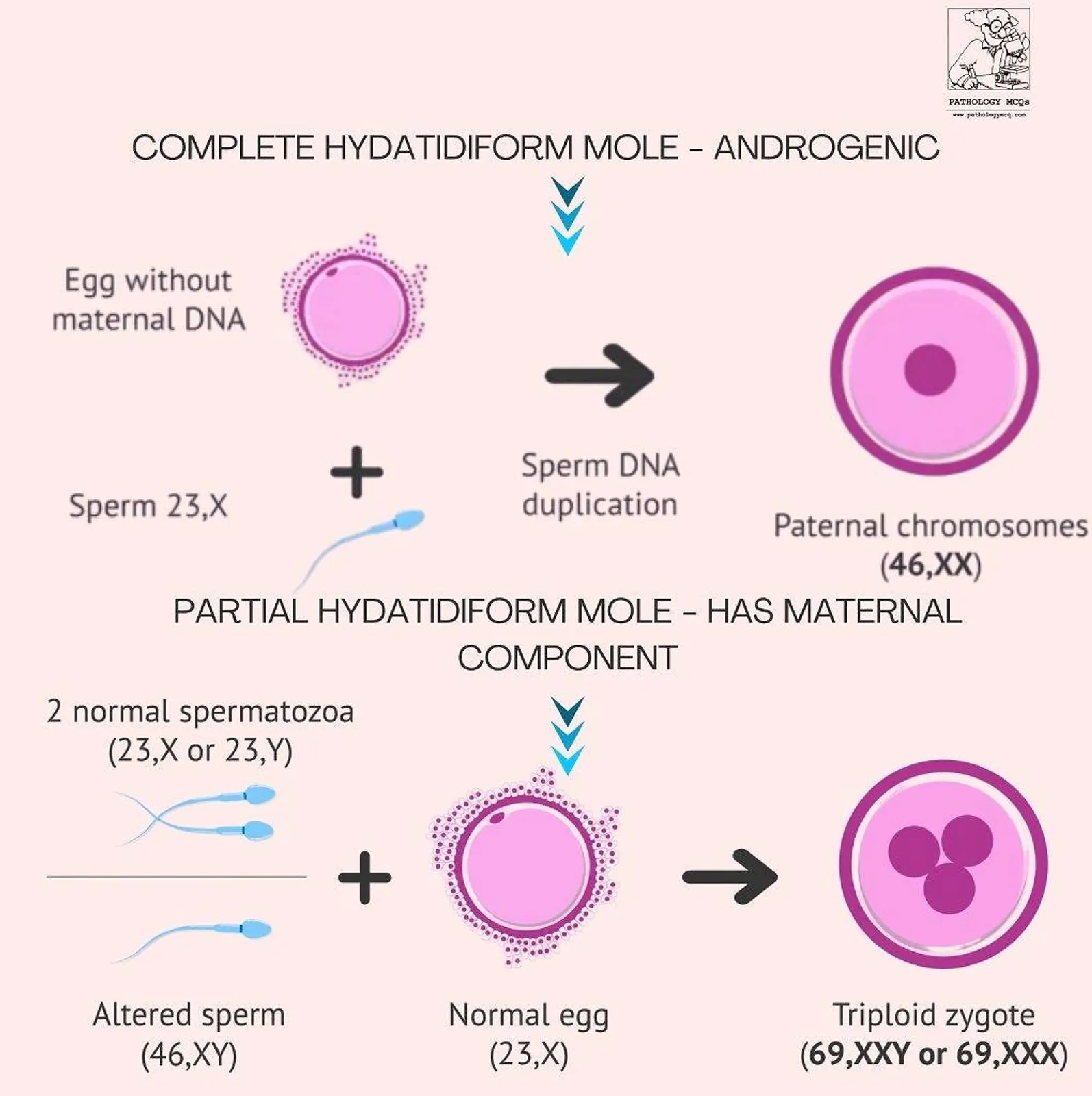
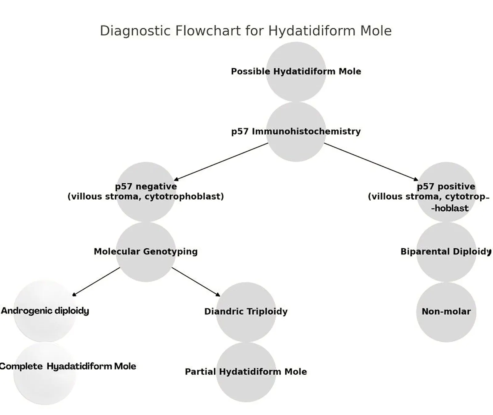
- Complete mole: Derived entirely from paternal DNA (most commonly 46,XX by duplication of sperm genome in an empty ovum), lacks maternal genome, and thus p57 is absent.
- Partial mole: Has a maternal component (usually triploid—2 paternal + 1 maternal), so p57 is expressed.
Histological Features of Hydatidiform Moles
Complete Hydatidiform Mole (CHM)
Key histopathologic features:
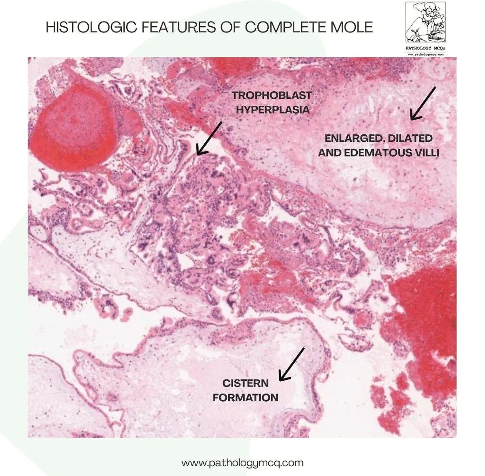
- Cistern formation – large central cavitations in villi.
- Diffuse trophoblastic hyperplasia – proliferation of cytotrophoblast and syncytiotrophoblast.
- Enlarged, edematous, avascular villi – with characteristic scalloping or irregular outlines.
Genetic basis: Androgenetic diploidy (46,XX or 46,XY).
VIEW THE INTERACTIVE CASE - TRY TO NOTE THESE HISTOLOGIC FINDINGS
Partial Hydatidiform Mole (PHM)
Key features:
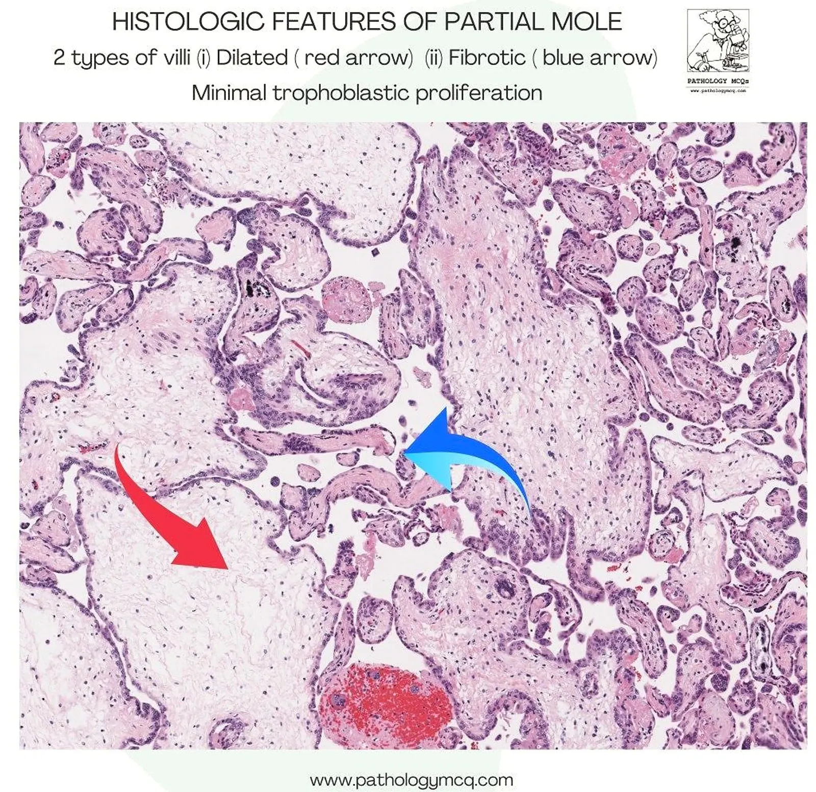
- Two populations of villi:
- Dilated villi (with cisterns)
- Fibrotic villi (shrunken with dense stroma)
- Mild or focal trophoblastic proliferation
- Fetal tissue may be present
Genetic basis: Triploidy (69,XXY or 69,XXX or 69,XYY) – includes maternal DNA.
p57 Immunohistochemistry: Diagnostic Application
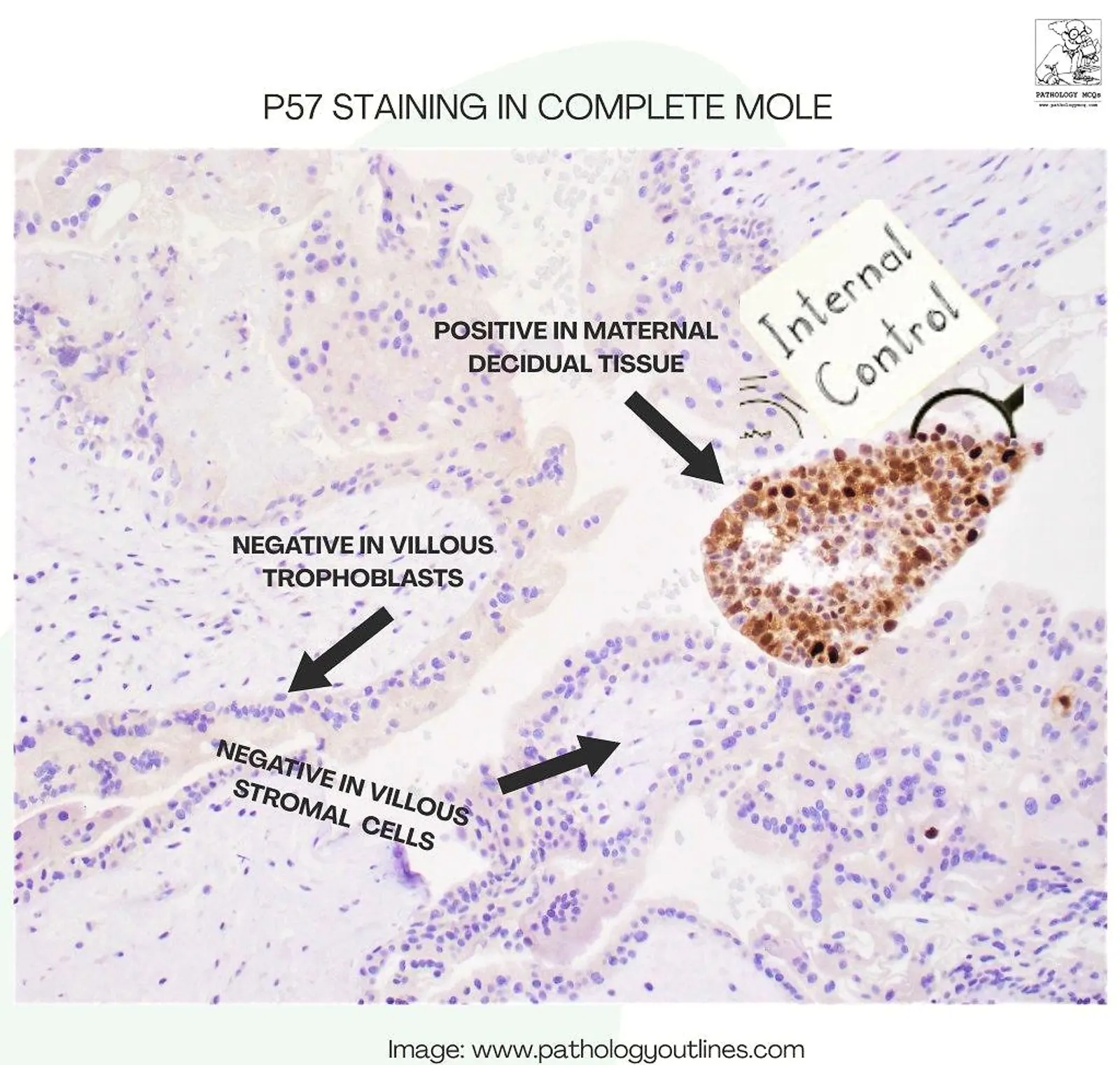
Complete Mole – p57 Negative
- Villous trophoblast: Negative
- Villous stromal cells: Negative
- Maternal decidua: Positive (internal positive control)
- Intermediate trophoblasts are also positive acting as an internal control.
This pattern confirms the absence of maternal genetic material in villous tissues.
🔍 Partial Mole – p57 Positive
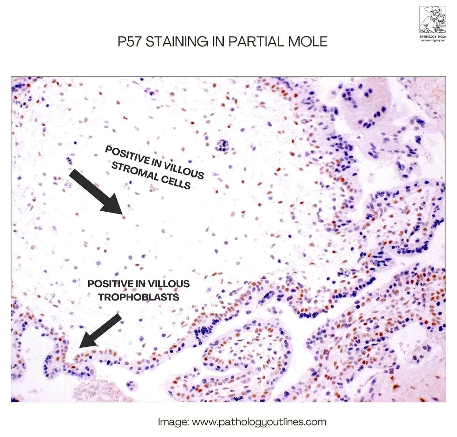
- Villous trophoblast: Positive
- Villous stromal cells: Positive
- Maternal decidua: Positive
The presence of maternal genetic material results in widespread p57 expression.
Summary Table: Interpreting p57 Expression
| Tissue Type | Complete Mole | Partial Mole | Non-molar Hydropic Pregnancy |
|---|---|---|---|
| Villous Trophoblast | Negative | Positive | Positive |
| Villous Stromal Cells | Negative | Positive | Positive |
| Decidual Tissue | Positive | Positive | Positive |

Pitfalls in Diagnosis Using p57
Despite its utility, p57 interpretation must be done with caution, considering potential diagnostic pitfalls:
1. Non-Molar Hydropic Abortions with Trisomies
- Trisomy 13, 18, or 21 can show hydropic villi but have normal biparental inheritance.
- p57 will be positive in these, potentially mimicking partial moles morphologically.
2. Misinterpretation Due to Sampling Error
- Focal or weak staining due to technical issues can be confused with complete mole.
- Always correlate with internal positive control (decidua).
3. Hybrid/Chimeric Pregnancies
- Rare cases may show a mix of androgenetic and biparental cell lines.
- May exhibit mosaic or hybrid staining patterns – some villi positive, others negative.
- These require molecular genotyping for definitive classification.
4. Early Gestational Age
- Early moles, especially before 8 weeks, may not show full-blown histologic features.
- p57 may still be helpful but must be interpreted in the right clinical context.
5. Extragenital Tissues Mistaken for Decidua
- Decidua must be identified correctly; myometrium or blood clots do not act as reliable internal controls.
Clinical Relevance
- p57 IHC is critical for differentiating a complete mole from partial mole and hydropic abortion when histology alone is inconclusive.
- Early diagnosis ensures prompt monitoring of β-hCG levels post-evacuation, as complete moles have a higher risk of progression to gestational trophoblastic neoplasia (GTN).
Reference
Kurman RJ, Ellenson LH, Ronnett BM. Blaustein's Pathology of the Female Genital Tract. 7th edition. Springer; 2019.
Banet N, DeScipio C, Murphy KM, Beierl K, Adams E, Vang R, Ronnett BM.
Characteristics of hydatidiform moles: Analysis of a prospective series with p57 immunohistochemistry and molecular genotyping
Modern Pathology. 2014;27:238–254.