
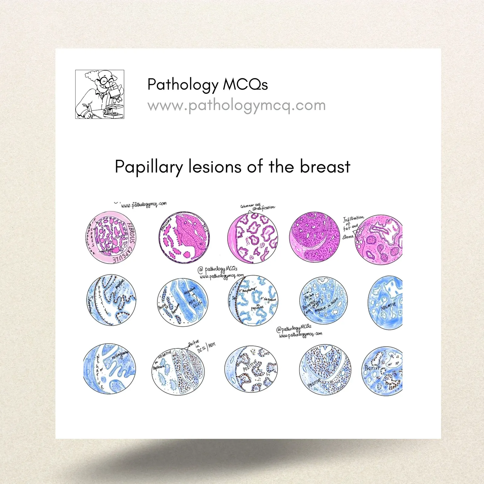
Papillary breast lesions present a diagnostic challenge due to their diverse morphologies and potential for malignancy. This blog post aims to summarize the key histopathological features of various papillary breast lesions, providing a helpful guide for pathology students, residents, and practicing pathologists.
Try these MCQs and test your knowledge before moving on.
Summary of Papillary Breast Lesions
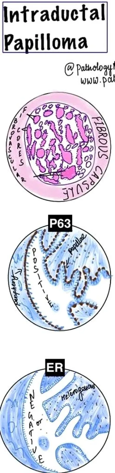
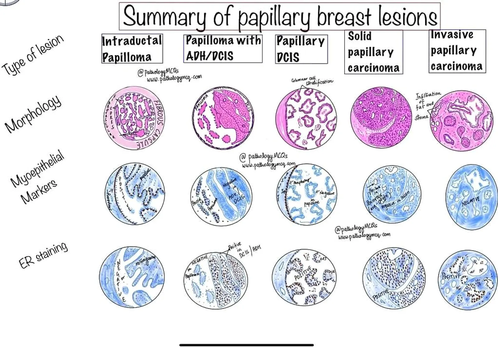
Intraductal Papilloma:
- Morphology: Characterized by a fibrovascular core, intraductal papillomas are benign lesions that typically do not show atypia.
- Myoepithelial Markers: These markers are positive, indicating the presence of myoepithelial cells throughout the lesion.
- ER Staining: Negative staining for estrogen receptors is common in these benign lesions.
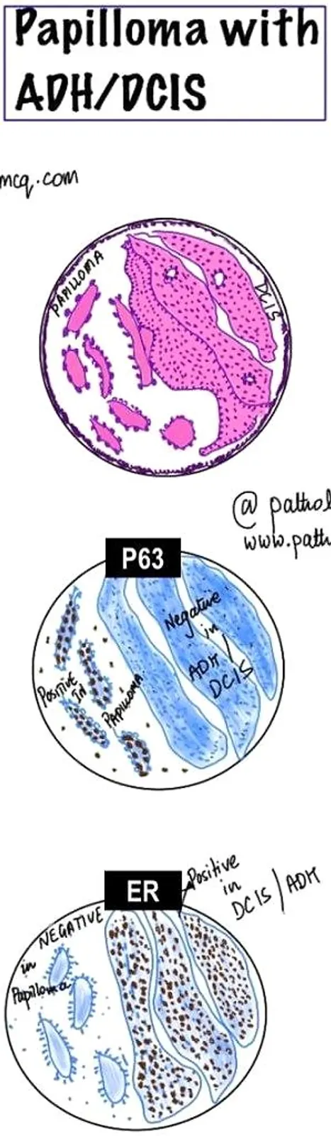
Papilloma with ADH/DCIS:
- Morphology: The lesion shows areas of atypical ductal hyperplasia (ADH) or ductal carcinoma in situ (DCIS) alongside benign papillomatous architecture.
- Myoepithelial Markers: There is partial or incomplete staining, particularly in the atypical areas. Positive staining is noted in the areas of papilloma.
- ER Staining: ER positivity is noted, especially in the ADH/DCIS components.
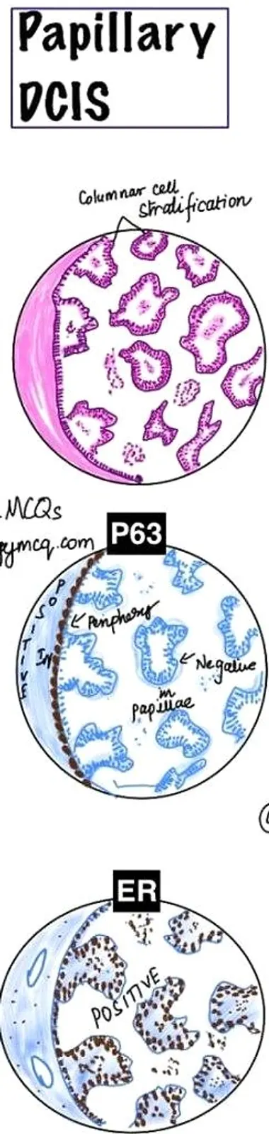
Papillary DCIS:
- Morphology: Exhibits glandular cell stratification without invasion.
- Myoepithelial Markers: Staining is absent in the central parts of the lesion, consistent with a diagnosis of DCIS.
- ER Staining: Strongly positive, reflecting the proliferative nature of the lesion.
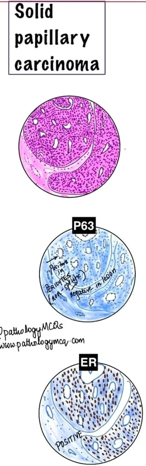
- Solid Papillary Carcinoma:
- Morphology: A solid pattern
- Myoepithelial Markers: Basal keratins may be variably positive in solid papillary carcinomas at the periphery of the lesion.
- ER Staining: Positive in most cases. Neuroendocrine markers can be noted in some cases.
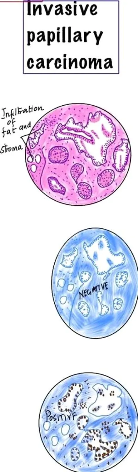
- Invasive Papillary Carcinoma:
- Morphology: A solid pattern with infiltration into the surrounding fat and stroma.
- Myoepithelial Markers: Often absent, aligning with its invasive characteristics.
- ER Staining: Positive in most cases, correlating with its carcinoma status.
- Encapsulated Papillary Carcinoma:
- Morphology: Well-circumscribed lesion, often with a thick fibrous capsule, and shows a cribriform pattern of growth.
- Myoepithelial Markers: Absent within the lesion but may be present at the periphery or capsule.
- ER Staining: Strongly positive
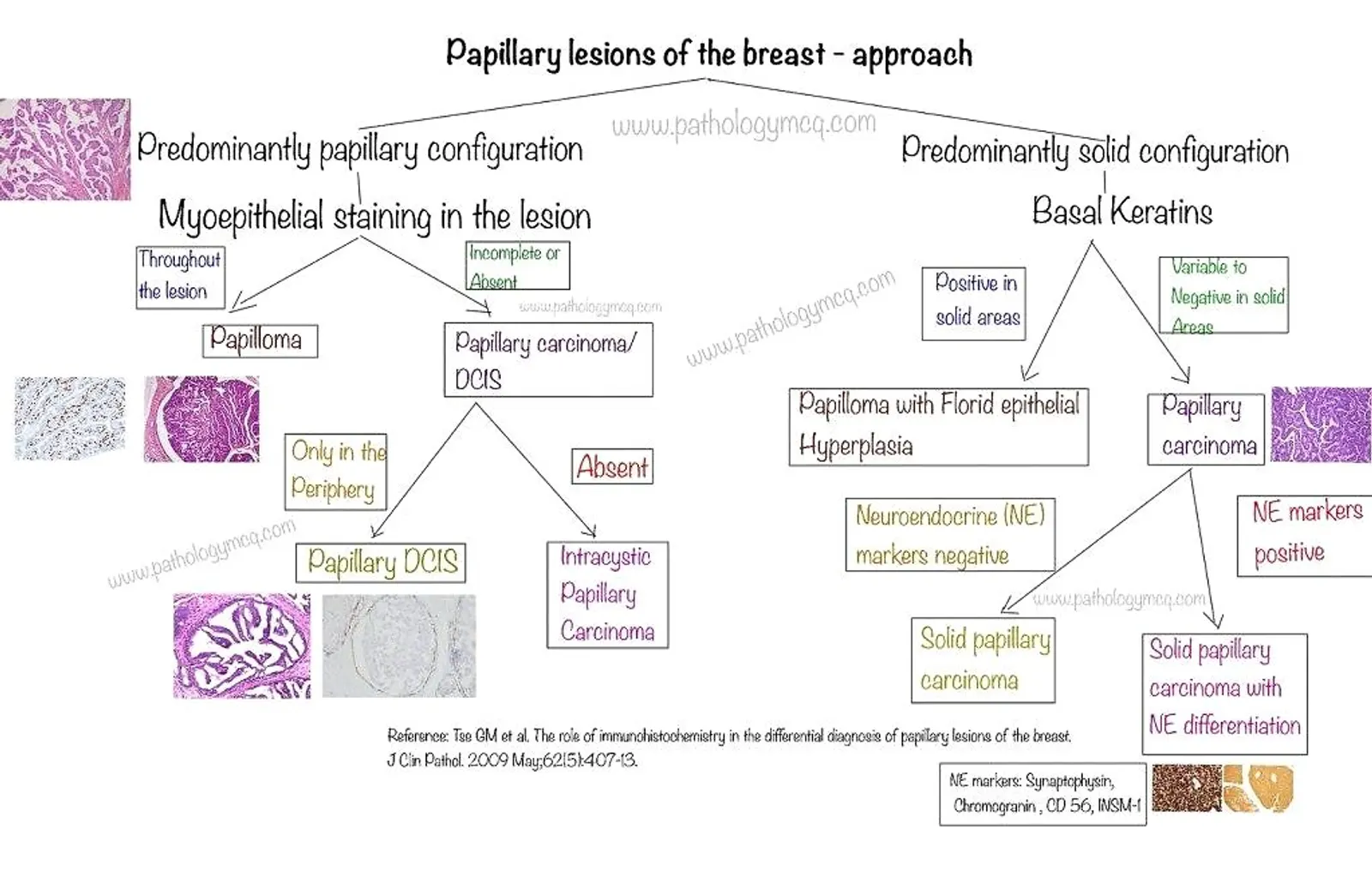
Approach to Diagnosing Papillary Lesions
The second image provides a structured approach to diagnosing papillary lesions of the breast. The decision tree is based on the presence of myoepithelial staining and the configuration of the lesion.
- Predominantly Papillary Configuration:
- Myoepithelial Staining Throughout the Lesion: This suggests a diagnosis of papilloma, a benign entity.
- Incomplete or Absent Myoepithelial Staining: Points towards papillary carcinoma or DCIS, depending on the lesion's periphery and myoepithelial marker status.
- Predominantly Solid Configuration:
- Basal Keratins Positive in Solid Areas: Suggests a benign papilloma with florid epithelial hyperplasia.
- Variable to Negative Basal Keratins in Solid Areas: Indicates a solid papillary carcinoma or carcinoma with neuroendocrine differentiation, necessitating further neuroendocrine marker testing.
NOTE: Basal keratins may be variably positive in solid papillary carcinomas at the periphery of the lesion.
Summary
| Lesion Type | Morphology | Myoepithelial Markers | ER Staining |
|---|---|---|---|
| Intraductal Papilloma | Fibrovascular core, benign, no atypia | Positive throughout the lesion | Negative |
| Papilloma with ADH/DCIS | Benign papilloma with areas of atypical ductal hyperplasia (ADH) or DCIS | Partially positive, particularly in benign areas | Positive, especially in ADH/DCIS areas |
| Papillary DCIS | Glandular cell stratification, no invasion | Absent in central lesion areas | Strongly positive |
| Solid Papillary Carcinoma | Solid pattern, infiltrative, involves fat and stroma | Often absent | Positive |
| Invasive Papillary Carcinoma | Invasive features, loss of ductal architecture | Negative | Positive |
| Encapsulated Papillary Carcinoma | Well-circumscribed, thick fibrous capsule, cribriform growth pattern | Absent within lesion, may be present at periphery or capsule | Strongly positive |
This table condenses the key histopathological features, making it easier to compare and contrast the different types of papillary breast lesions.
ANSWER A FEW QUESTIONS TO TEST YOUR KNOWLEDGE
For more like this- JOIN US
Useful for - NEET SS, FRCPath, DHA and anatomic pathology boards (APCP Boards).