
Invasive fungal infections are life-threatening conditions caused by fungi that invade deep tissues, often in immunocompromised patients. Understanding their histopathology, clinical features, and treatment is essential for medical students, pathologists, and clinicians.
Subscribe to receive more such posts
Causes & Risk Factors:
- Immunosuppression – chemotherapy, corticosteroids, or transplant immunosuppressants.
- Diabetes mellitus (especially ketoacidosis) – predisposes to mucormycosis.
- Prolonged ICU stay – invasive devices, broad-spectrum antibiotics, and catheters increase risk of Candida infections.
- Environmental exposure – inhalation of spores from soil, bird droppings, and decaying matter (Histoplasma, Coccidioides).
- Disrupted normal flora – allows Candida overgrowth.
Symptoms:
- Pulmonary involvement – fever, persistent cough, hemoptysis, pleuritic chest pain.
- CNS involvement – headache, confusion, seizures (notable in cryptococcosis).
- Sinonasal mucormycosis – nasal obstruction, facial pain, black necrotic eschar.
- Disseminated candidiasis – persistent fever, skin lesions, multi-organ dysfunction.
Diagnosis:
Narrow-based budding:
- Very small organisms (2–6 µm).
- Appear as round yeast with narrow stalk-like budding.
- In Cryptococcus, a clear halo (capsule) is seen around the yeast in H&E stains.
- In Histoplasma – 2–6 μm, narrow-based budding, intracellular (within macrophages), halo appearance.
- Stains: H&E (shows halo), Silver stain (better contrast).

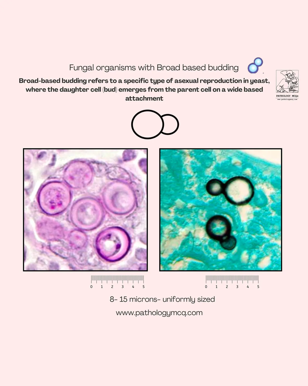
Broad-Based Budding Yeasts:
- Larger organisms (8–15 µm).
- Yeast cells are uniform in size.
- Daughter cell forms with a broad base of attachment (compared to narrow budding in cryptococcus).
- Thick, double-walled yeast.
Spherules with Endospores:
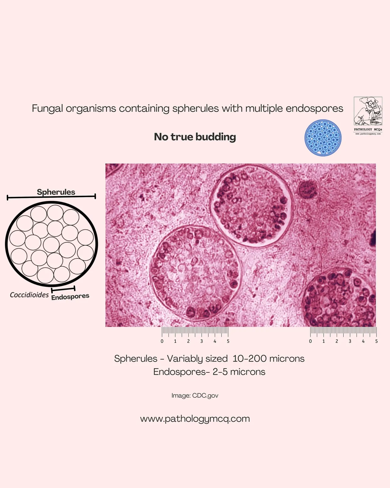
Coccidioides
- Large spherules (10–200 µm) containing smaller endospores (2–5 µm).
- No true budding.
- Seen in tissue biopsies of coccidioidomycosis.
- Important differentiator from budding yeasts.
Multiple Budding Yeast:
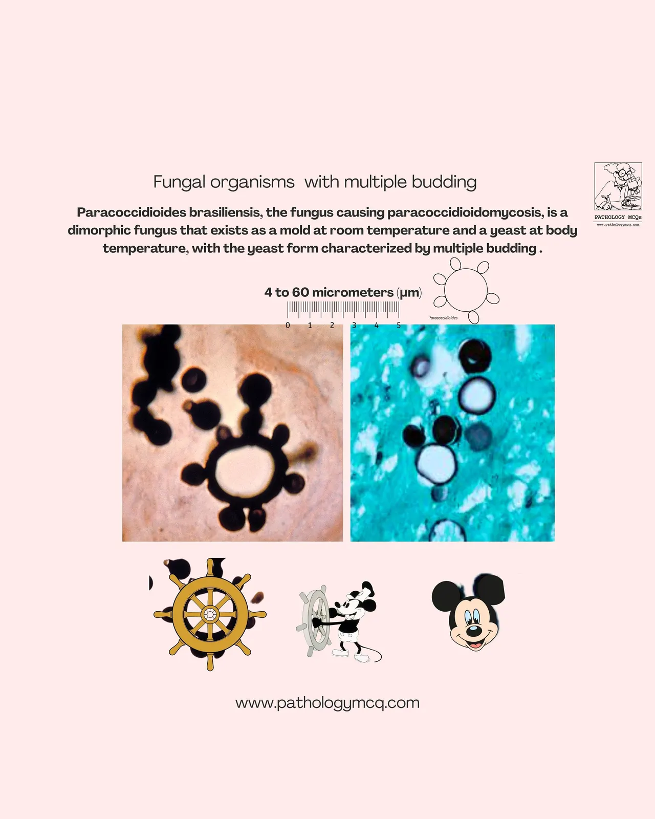
Paracoccidioides
- Variable size (4–60 µm).
- Characteristic “pilot wheel” or “mariner’s wheel” appearance.
- Multiple buds arise simultaneously from the mother yeast cell.
- Dimorphic fungus: mold at room temp, yeast at body temp.
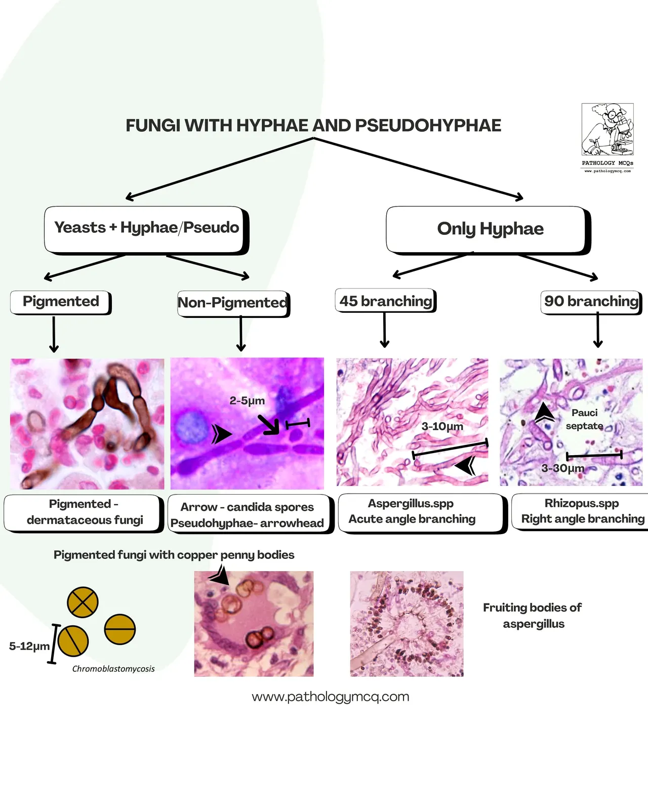
Fungi with Hyphae and Pseudohyphae:
Candida
- Yeast + pseudohyphae seen.
- Pseudohyphae appear as elongated budding cells, not true hyphae
- Important in mucosal infections.
Aspergillus
- Thin septate hyphae with acute angle branching (45°).
- Fruiting bodies occasionally seen.
- Common cause of invasive pulmonary infections.
Rhizopus
- Broad, non-septate hyphae with right angle branching (90°).
- Seen in diabetic ketoacidosis and immunosuppressed patients.
Treatment:
- Candida – Echinocandins (first line), fluconazole if susceptible.
- Aspergillus – Voriconazole (drug of choice).
- Mucormycosis – Liposomal Amphotericin B + surgical debridement.
- Cryptococcus (meningitis) – Amphotericin B + Flucytosine, followed by fluconazole.
- Histoplasmosis / Coccidioidomycosis / Paracoccidioidomycosis – Itraconazole for mild-to-moderate disease, Amphotericin B for severe cases.
- Supportive measures: control diabetes, reduce immunosuppressants if possible, and remove infected catheters.
Prevention:
- High-risk patients may receive antifungal prophylaxis (e.g., posaconazole in transplant patients).
- Hospital infection control – HEPA filters, minimizing invasive devices.
- Lifestyle precautions – immunocompromised patients should avoid construction sites, soil, and bird droppings.
- Glycemic control – reduces mucormycosis risk.
- Judicious antibiotic use – prevents fungal overgrowth.
Mnemonics:
| Fungus | Morphology | Histology Feature |
| Cryptococcus | Narrow budding | Halo, capsule |
| Blastomyces | Broad budding | Uniform yeast |
| Coccidioides | Spherules | Endospores inside |
| Paracoccidioides | Multiple budding | Pilot wheel |
| Aspergillus | Hyphae | 45° branching |
| Rhizopus | Hyphae | 90° branching |
- ABC-P for budding fungi: Aspergillus (acute angle), Blastomyces (broad), Cryptococcus (capsule, narrow), Paracoccidioides (pilot wheel).
Multiple choice questions (MCQs):
Invasive Fungal Infections — 5 MCQ Challenge
0/5 answered · Score: 0
⏳02:00
Your Result
Time to test what you have learnt
Identify this fungus?
Check answer
CORRECT ANSWER IS MUCOR
[openseadragon_viewer dzi="2025/08/c4/c4" regions="2025/08/c4/reg.json"]Conclusion:
Invasive fungal infections remain a critical challenge in pathology, requiring accurate histopathological recognition for timely diagnosis. Early identification and treatment not only improve patient outcomes but also strengthen exam preparation for aspiring pathologists.