
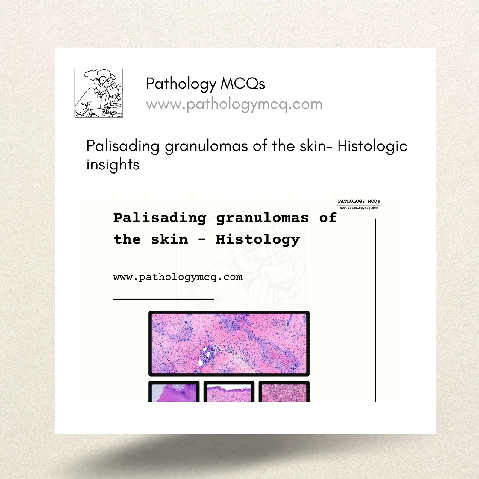
Palisading granulomas are a fascinating histopathological feature encountered in dermatopathology. They are characterized by histiocytes arranged in a palisading pattern around areas of necrosis or altered connective tissue. This article will guide you through the key types of palisading granulomas and their histological features.
Understanding Palisading Granulomas
Palisading granulomas are broadly divided into two types based on their histological characteristics:
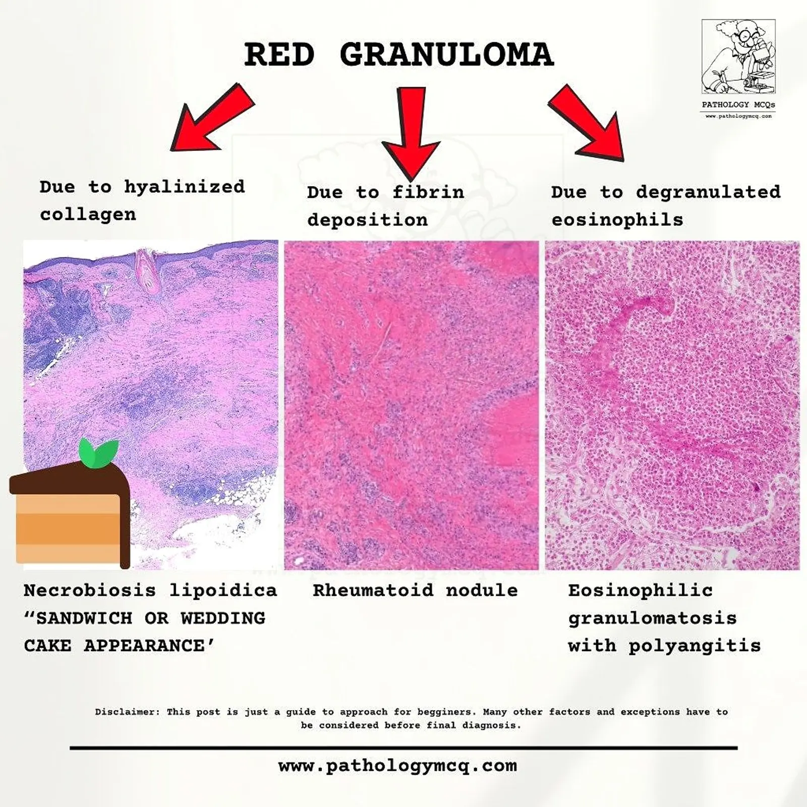
Red Granulomas
These granulomas are associated with fibrin deposition, hyalinized collagen, or degranulated eosinophils.
- Examples:
- Necrobiosis lipoidica: Characterized by "sandwich or wedding cake" appearance due to layers of necrobiosis and inflammation.
- Rheumatoid nodule: Shows fibrinoid necrosis surrounded by palisading histiocytes.
- Eosinophilic granulomatosis with polyangiitis: Features eosinophils and areas of necrosis.
Blue Granulomas
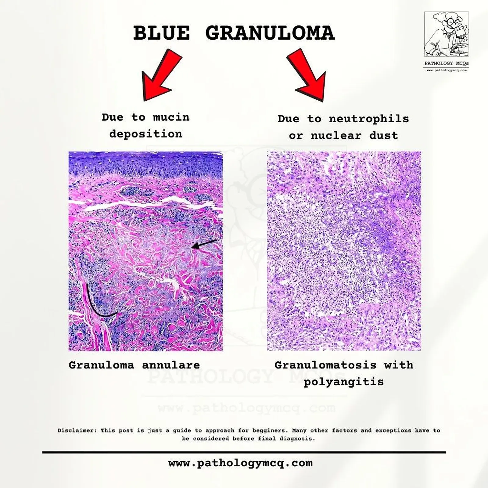
These granulomas involve mucin deposition or nuclear dust.
- Examples:
- Granuloma annulare: Exhibits mucin deposition in the dermis with histiocytes forming a palisading arrangement.
- Granulomatosis with polyangiitis: Shows neutrophils and nuclear debris in a palisading pattern.
Comparison of Red and Blue Granulomas
| Feature | Red Granuloma | Blue Granuloma |
|---|---|---|
| Key Histological Feature | Hyalinized collagen, fibrin, eosinophils | Mucin deposition, nuclear dust |
| Examples | Necrobiosis lipoidica, Rheumatoid nodule | Granuloma annulare, Granulomatosis |
| Clinical Relevance | Associated with systemic diseases | Often confined to localized conditions |
Histological Pearls for Palisading Granulomas
- Necrobiosis lipoidica: Look for thickened collagen with a layered inflammatory response.
- Rheumatoid nodule: Central necrotic zone surrounded by palisading macrophages and lymphocytes.
- Granuloma annulare: Abundant mucin between collagen bundles and a sparse inflammatory infiltrate.
Helpful Mnemonics
- Red: "Red FLAGS"
- Blue: "Blue MAN"

Interactive video explanation
Conclusion
Palisading granulomas are a hallmark of various dermatological and systemic conditions. Understanding their histological features and clinical context is essential for accurate diagnosis and management. For more detailed notes, Case based interactive discussions and case based MCQs, visit